What is a Jones fracture?
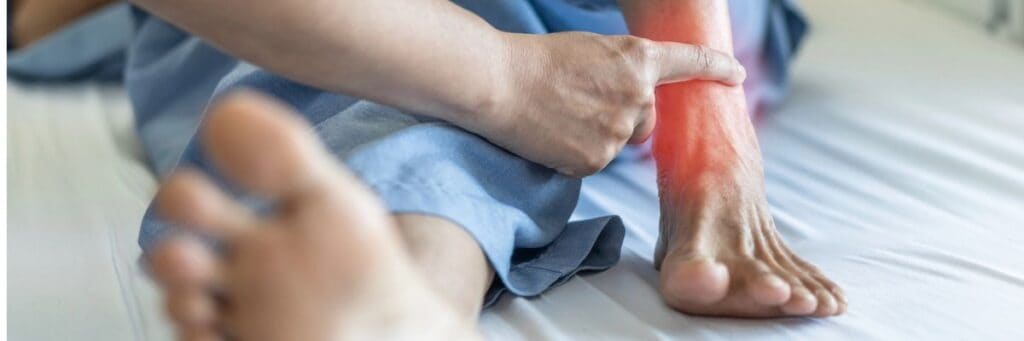
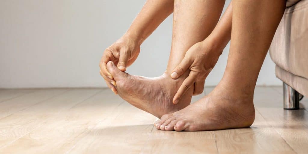
A Jones fracture is a break at the base of the fifth metatarsal bone, which is the long bone on the outside of the foot leading to the pinky toe. This common foot injury frequently affects both elite athletes and weekend warriors.
This area has a poor blood supply, which makes healing difficult. Because a Jones fracture is very similar to a sprain or avulsion fracture (known as a “pseudo-Jones” injury), an experienced orthopedic sports medicine foot and ankle specialist should evaluate it.
A Jones fracture is a break in the fifth metatarsal, the long bone on the outer edge of your foot that connects your little toe to your heel. Specifically, it occurs in the diaphysis, a narrow section of the bone’s shaft where blood supply is naturally limited. That poor circulation is what makes Jones fractures notoriously slow to heal and prone to complications like non-union and delayed union. Named after orthopedic surgeon Sir Robert Jones, who described the injury in 1902, this fracture is one of the most common foot injuries among athletes and active adults.
Anatomy and fracture zones of the fifth metatarsal
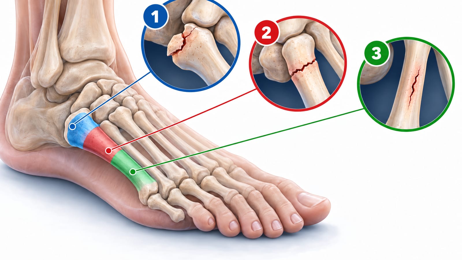
Where your fracture sits on the fifth metatarsal matters greatly for treatment and prognosis. Specialists classify fifth metatarsal fractures into three zones:
Zone 1 – Avulsion fracture (pseudo-Jones fracture): A break at the tuberosity, the bony bump at the very base of the fifth metatarsal. The peroneus brevis tendon or lateral ligament pulls a small bone fragment away during a sudden ankle roll. This is also called a dancer’s fracture. Healing typically takes 3-12 weeks with conservative care.
Zone 2 – True Jones fracture: A break at the junction between the tuberosity and the diaphysis. This zone has the poorest blood supply of the three, making it the most difficult to heal and the most likely to result in non-union or refracture. Studies report refracture rates as high as 50% with non-surgical management alone.
Zone 3 – Stress fracture: A fracture further down the diaphysis, usually caused by repetitive stress rather than a single acute trauma. Common in distance runners and basketball players, Zone 3 injuries carry a high risk of delayed union and often require surgical intervention.
The avulsion fracture and the true Jones fracture are frequently confused because both cause pain on the outer foot after an ankle roll. The key distinction is location: Zone 1 is at the tuberosity and generally heals well; Zone 2 is at the diaphysis and requires more aggressive treatment. An X-ray, and sometimes an MRI or CT scan, is needed to tell them apart definitively.
What are the symptoms of a Jones fracture?
Typical symptoms include:
Sharp or aching pain along the outer edge of the foot
Swelling and bruising near the base of the little toe
Difficulty bearing weight or walking with a noticeable limp
Tenderness directly over the fifth metatarsal when pressed
Some patients recall the exact moment of injury, often a pop during a lateral cut or ankle roll. Others, particularly those with stress fractures, describe a dull ache that has built gradually over weeks. Either way, symptoms that persist beyond a few days warrant prompt evaluation.
Where exactly you feel pain matters, because the fifth metatarsal is divided into three distinct zones – and each zone behaves differently. Zone 1 sits at the very tip of the bone’s base and is the site of avulsion fractures (sometimes called pseudo-Jones fractures), where a tendon or ligament yanks a small fragment away during an ankle roll. Zone 2 is the narrow, blood-poor junction just beyond that – the true Jones fracture zone – and it heals slowly because the local blood supply is limited. Zone 3 extends further down the shaft and is where repetitive stress fractures tend to develop, often without a single traumatic event. Knowing which zone is involved helps Dr. Baravarian build the right tailored treatment plan from the start.
Higher-risk groups include elite athletes, dancers, weekend warriors, employees who stand all day, and patients with osteoporosis or a high-arched foot, since a cavus foot places extra load on the outer metatarsal with every step. Sudden changes in training volume, hard court surfaces, and worn-out footwear are also common contributing factors – particularly in Zone 3 stress fractures, where the bone is weakened by cumulative load rather than a single misstep. If you recognize your symptoms in any of these patterns, early imaging and a personalized evaluation are the fastest path to an accurate diagnosis and faster healing.
What causes a Jones fracture?
Now that you know what a Jones fracture feels like, it helps to understand how one actually happens – because it’s not always as dramatic as you might expect. There are two main pathways: acute trauma and repetitive stress.
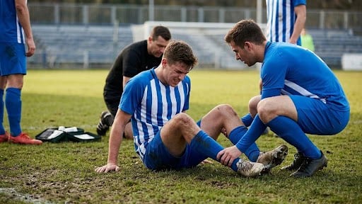
Acute trauma is the more obvious culprit. A sudden, forceful movement – like rolling your ankle outward, planting your foot and cutting sharply to one side, or landing awkwardly after a jump – can place enormous stress on the fifth metatarsal in an instant. This is why Jones fractures are so common among basketball players, soccer players, and dancers. If you’ve ever watched an athlete go down after a quick change of direction, there’s a good chance a Jones fracture was involved.
Repetitive stress is the sneakier cause. Over time, repeated impact and loading of the outer foot, common in runners and high-mileage athletes, can gradually weaken the bone until it finally gives way, even without a single dramatic incident.
Certain factors also raise your risk. A high-arched foot (cavus foot) shifts more weight to the outer edge of the foot, increasing pressure on the fifth metatarsal with every step. Overpronation, poor footwear, and returning to activity too quickly after a previous injury can also contribute. Understanding your personal risk factors is key to building a tailored treatment plan that works long term.
How is a Jones fracture diagnosed?
Getting an accurate diagnosis is one of the most important steps in treating a Jones fracture – and it’s not always as straightforward as it sounds. Dr. Bob and his team use a combination of physical examination and state-of-the-art imaging to ensure they don’t miss anything.
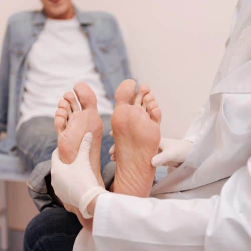
The process typically starts with a thorough physical exam. Dr. Bob will gently press along the outer edge of your foot, checking for tenderness directly over the fifth metatarsal. He’ll also assess swelling, bruising, and how well you can bear weight. This hands-on, personalized care approach helps pinpoint the injury before a single image is taken.
Next comes imaging – and this is where things get nuanced. X-rays are the standard first step and can confirm most Jones fractures. However, here’s an important caveat: stress fractures in Zone 3 and even some Zone 2 fractures may not show up on initial X-rays, especially in the first week or two after injury. If your symptoms strongly suggest a fracture but the X-ray looks normal, don’t be discouraged – that doesn’t mean you’re in the clear.
In those cases, Dr. Bob may recommend an MRI or CT scan. An MRI is particularly sensitive for detecting early stress fractures and bone marrow changes that X-rays simply can’t capture. A CT scan, on the other hand, gives a detailed look at bone structure and is especially useful for surgical planning. If you’re a runner or athlete dealing with recurring outer foot pain, learning more about common foot problems for runners can also help you understand your risk factors before a fracture fully develops.
What are the non-surgical Jones fracture treatment options?
For many patients, especially those with Zone 1 avulsion fractures or non-displaced Zone 2 fractures, non-surgical treatment is appropriate and includes:
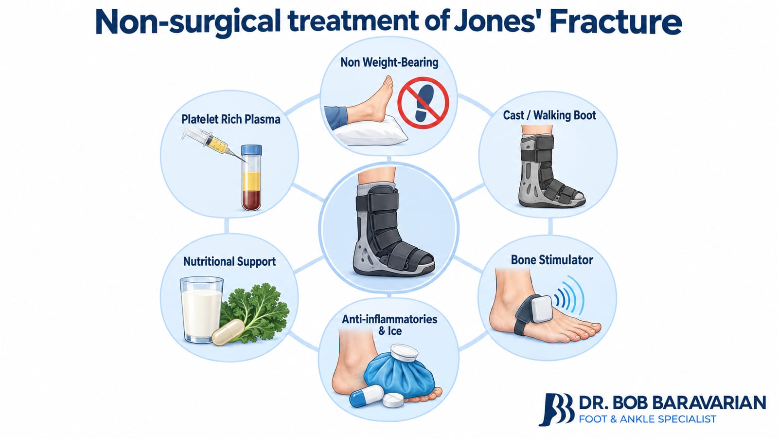
Non-weight bearing: Crutches for the first 4-6 weeks to protect the fracture site from compressive load. Because the Jones fracture zone sits in a naturally low-blood-supply area, keeping all weight off the foot during this window is non-negotiable – even brief unprotected steps can displace the fracture and push you toward surgery. Dr. Bob typically reassesses weight-bearing status at the four-week mark using follow-up X-rays before allowing any transition to partial weight-bearing in a boot.
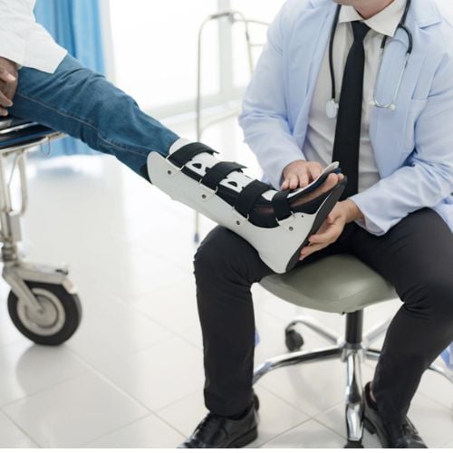
Cast immobilization or walking boot: A short-leg cast or rigid boot keeps the bone aligned and reduces the risk of refracture. Most patients transition from a cast to a removable walking boot around weeks four to six, depending on how well the fracture is consolidating on imaging. The boot allows controlled, protected weight-bearing while still shielding the healing bone from rotational stress.
Bone stimulator: Low-intensity pulsed ultrasound or electrical stimulation devices can accelerate bone healing, particularly useful for delayed unions. Dr. Bob recommends bone stimulator therapy early in the treatment course for true Jones fractures – not just as a rescue tool when healing stalls. Peer-reviewed studies show that daily 20-minute sessions meaningfully shorten time to union, which matters enormously in a zone already prone to slow healing. Athletes and active patients especially benefit from starting stimulation in the first two weeks.
Anti-inflammatory medication and icing: Reduce acute swelling and pain; ice for 15-20 minutes several times daily. NSAIDs are used judiciously in the early acute phase – prolonged use can actually interfere with bone healing, so Dr. Bob typically limits anti-inflammatory medication to the first one to two weeks and transitions patients to other pain management strategies as healing progresses.
Nutritional support: Adequate vitamin D and calcium intake are essential for bone repair; Dr. Bob evaluates levels and recommends supplementation when needed. Vitamin D deficiency is surprisingly common and is a frequently overlooked reason why Jones fractures fail to heal on schedule. Dr. Bob routinely checks serum vitamin D and calcium levels at the initial consultation. Patients with low levels are placed on a targeted supplementation protocol – typically vitamin D3 combined with magnesium for optimal absorption – because getting these numbers into the optimal range can make a measurable difference in how quickly the bone knits together.
Platelet-rich plasma (PRP): As part of his regenerative approach, Dr. Bob may use PRP injections to concentrate the body’s own growth factors at the fracture site, promoting faster, more robust healing. PRP is drawn from a small sample of your own blood, spun in a centrifuge to concentrate the platelets, and then precisely injected at the fracture site under imaging guidance. The growth factors released by those platelets – including PDGF and TGF-β – directly stimulate bone cell activity and new blood vessel formation, addressing the poor vascularity that makes Jones fractures so stubborn in the first place. For patients who want every advantage in their recovery, PRP is a powerful addition to the non-surgical toolkit that Dr. Bob has offered his patients for years.
Healing typically takes 6-8 weeks for most fractures, but true Jones fractures often require closer to four to five months. Studies report non-union or refracture in up to 50% of conservatively managed cases, which is why close follow-up imaging is critical. If X-rays at the six-to-eight-week mark show insufficient callus formation, Dr. Bob will discuss whether continuing conservative care with added bone stimulation and PRP makes sense, or whether surgery would offer a faster, more reliable path back to full activity.
What are the surgical treatment options for a Jones fracture?
Surgery is recommended when the fracture is displaced, involves multiple fragments, has failed to heal with conservative care, or when the patient, particularly a competitive athlete, cannot afford a prolonged recovery. Surgical treatment dramatically shortens return-to-sport timelines compared to non-surgical management.
The standard procedure is intramedullary screw fixation: a cannulated screw is placed down the center of the fifth metatarsal to compress and stabilize the fracture. Screw diameter is a critical variable – research consistently shows that larger-diameter screws provide superior rotational stability and significantly reduce the risk of refracture, which is a real concern given the Jones fracture’s notoriously poor blood supply. Screw sizing matters, as a larger-diameter screw provides superior rotational stability and reduces refracture risk. When bone quality is poor or non-union is present, surgeons add bone grafting to stimulate new bone formation at the fracture site. Bone graft options range from autograft – harvested from the patient’s own body for maximum biological compatibility – to allograft and synthetic bone substitutes, each chosen based on the degree of non-union and the patient’s overall bone health.
Dr. Bob’s minimally invasive approach uses a small incision, preserving surrounding soft tissue and reducing post-operative pain. This state-of-the-art technique means less disruption to the delicate structures around the fifth metatarsal, translating directly into less pain and faster healing for his patients. He also incorporates PRP at the surgical site to accelerate biological healing. PRP (Platelet-Rich Plasma) is drawn from the patient’s own blood, concentrated, and applied directly to the fracture zone during surgery. The growth factors released by the platelets supercharge the body’s natural repair process, which is especially valuable at the Jones fracture site where blood flow is already limited. This regenerative, pioneering approach is a key part of Dr. Bob’s tailored treatment plan for every surgical patient. Post-operatively, patients follow a structured protocol progressing from non-weight bearing to a walking boot over 6-8 weeks, with follow-up X-rays confirming bone consolidation before resuming full activity.
Jones fracture recovery and return to sport
Recovery from a Jones fracture is a phased process that depends on whether you were treated conservatively or surgically, and how well the fracture site responds to healing. Because the Zone 2 true Jones fracture sits in a notoriously low-blood-supply area, patience and a structured rehabilitation plan are non-negotiable. Dr. Bob tailors every recovery plan to the individual patient, but the milestones below reflect what most patients can expect:
Weeks 1-6: Non-weight bearing in a cast or boot; manage pain and swelling; gentle range-of-motion exercises to prevent stiffness. Surgical patients – particularly those who underwent minimally invasive fixation – often move through this phase more comfortably, with less pain and swelling than patients treated with traditional open techniques.
Weeks 6-10: Progressive weight bearing in a walking boot as X-rays confirm healing; physical therapy begins, focusing on restoring range of motion and reducing scar tissue. If imaging shows the fracture line is consolidating well, your therapist will introduce gentle peroneal and intrinsic foot strengthening at this stage.
Weeks 10-14: Transition to a supportive shoe; strengthen the foot intrinsics, peroneals, and calf; add balance and proprioception training. Proprioception work is especially important here – the muscles around the fifth metatarsal are often deconditioned after weeks of immobilization, and rebuilding neuromuscular control dramatically lowers your refracture risk.
Weeks 14-20: Sport-specific conditioning, gradual return to running, cutting, and jumping under physical therapy supervision. Athletes returning to lateral-movement sports like basketball or soccer should expect a progressive loading program before full clearance – rushing this phase is the most common reason Jones fractures recur.
Return to full sport: Most surgical patients return to competitive sport between 3 and 5 months. Conservative cases may take 5-6 months, and some non-union cases require revision surgery if the fracture fails to heal. Clearance requires both clinical and imaging confirmation that the fracture has fully healed.
Physical therapy is a cornerstone of recovery. A structured program addresses the muscle weakness and altered gait mechanics that often develop during immobilization, reducing the risk of refracture once you return to activity. Dr. Bob also evaluates contributing biomechanical factors – such as a high-arch cavus foot or tight peroneal tendons – that may have predisposed you to the fracture in the first place, building those corrections into your tailored treatment plan so you come back stronger, not just healed.
Prognosis and why choosing the right specialist matters
With proper treatment, most Jones fracture patients return to full activity without long-term limitations. Surgical patients, particularly those treated with appropriately sized intramedullary screws and regenerative adjuncts like PRP, enjoy the best outcomes and the lowest refracture rates. A good prognosis depends on early, accurate diagnosis, correct zone classification, and a specialist who understands the nuances of fifth metatarsal biology.
Dr. Bob Baravarian is a trained trauma specialist and one of the nation’s foremost experts in foot and ankle fractures. Using pioneering minimally invasive and regenerative techniques, many of which he helped develop, he maintains among the highest Jones fracture success rates in the United States. He takes the time to get to know every patient personally, tailoring each treatment plan as if he were caring for his own family. His practice is conveniently located in Los Angeles, near Cedars-Sinai Medical Center, serving patients throughout Southern California. If you are dealing with outer foot pain after an injury or a gradual ache that won’t resolve, reach out to Dr. Bob’s office today and take the first step toward confident, expert care.
Jones fracture FAQs
What foods help broken bones heal faster?

While no magic food can mend a broken bone overnight, a balanced diet rich in certain nutrients can support healing. This includes foods rich in calcium, vitamin D, protein, vitamin C, vitamin K, omega-3 fatty acids, magnesium, and zinc.
How can you increase blood flow to a Jones fracture?
Increasing blood flow to a Jones fracture can support healing because an adequate blood supply delivers nutrients and oxygen to the injured area. Here are some general tips that may help:
- Rest and Immobilization
- Elevation
- Ice
- Compression
- Physical Therapy
- Nutrition: A balanced, nutrient-rich diet can support healing and improve blood circulation.
- Smoking Cessation
- Medications: Your doctor may prescribe medications to manage pain and inflammation, which can indirectly support blood flow by reducing discomfort.
How soon can I run after a 5th metatarsal fracture?
The timing for starting to run again after a 5th metatarsal fracture varies based on several factors, including fracture type, treatment, and bone-healing rate.
People heal at their own pace, and it’s essential to prioritize healing over the desire to return to running quickly. Rushing back into high-impact activities too soon can lead to reinjury or delayed healing.
How did Jones Fracture get its name?
The name Jones fracture comes from Sir Robert Jones, who described the fracture in Annals of Surgery in 1902. He discussed the findings of the fracture in 6 patients, including himself. He sustained a fifth metatarsal fracture after a night of dancing, where he noted that, aside from a traumatic injury, overloading and applying continued stress along the bone can lead to fracture.
Sources
-
Rosenberg GA, Sferra JJ. Treatment strategies for acute fractures and nonunions of the proximal fifth metatarsal. J Am Acad
https://pubmed.ncbi.nlm.nih.gov/11029561/ -
Torg JS, Balduini FC, Zelko RR, Pavlov H, Peff TC, Das M. Fractures of the base of the fifth metatarsal distal to the tuberosity. Classification and guidelines for non-surgical and surgical management.
https://pubmed.ncbi.nlm.nih.gov/6693447/ -
Chuckpaiwong B, Queen R, Easley M, Nunley J. Distinguishing Jones and Proximal Diaphyseal Fractures of the Fifth Metatarsal. Clin Orthop Relat Res. 2008;466(8):1966-70. doi:10.1007/s11999-008-0222-7
-
Bernstein DT, Mitchell RJ, McCulloch PC, Harris JD, Varner KE. Treatment of Proximal Fifth Metatarsal Fractures and Refractures With Plantar Plating in Elite Athletes. Foot Ankle Int.
https://pubmed.ncbi.nlm.nih.gov/30079768/ -
Mulcahey, M. K. (2021). Toe and forefoot fractures.
https://orthoinfo.aaos.org/en/diseases–conditions/toe-and-forefoot-fractures