What is an osteochondral lesion of the talus?
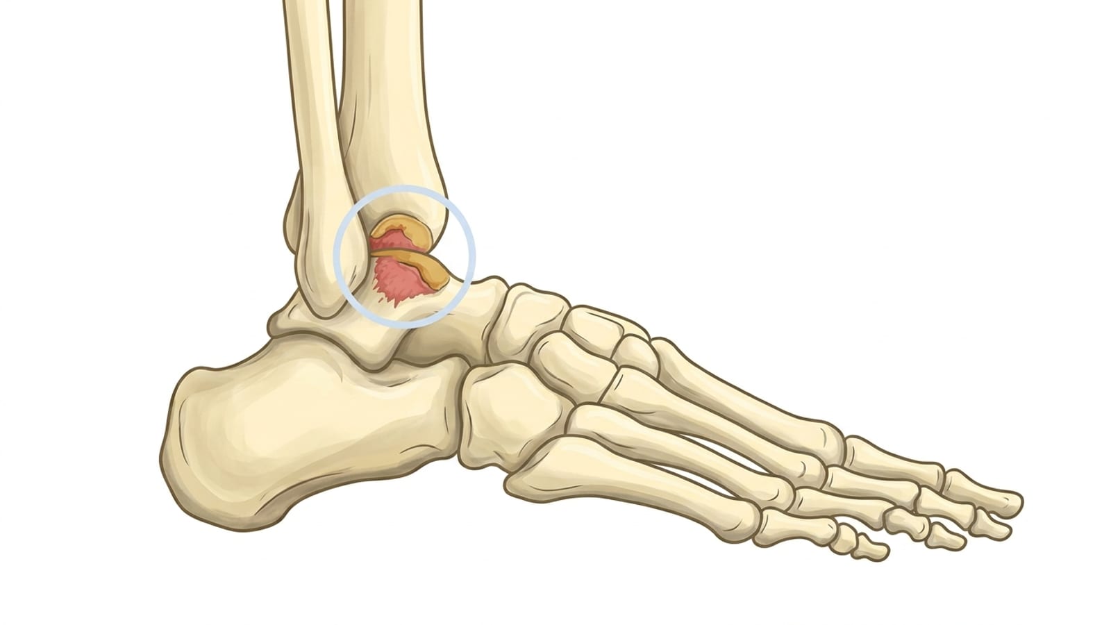
An osteochondral lesion of the talus (OLT) is an injury to the articular cartilage on the surface of the talus bone and the subchondral bone directly beneath it. The talus sits at the base of the shin bone (tibia) and calf bone (fibula), forming the lower half of the ankle joint. Because the entire top surface of the talar dome is covered by smooth hyaline cartilage, even a small area of damage can cause persistent pain, swelling, and mechanical symptoms that significantly affect daily life.
It is important to distinguish between two related but different injury types. A chondral lesion involves damage to the cartilage layer only, while a true osteochondral lesion (also called an osteochondral defect, or OCD) extends into the underlying bone. When the subchondral bone is involved, healing becomes more complex because bone and cartilage have very different repair capacities. Cartilage, in particular, has a very limited blood supply, which means it cannot regenerate on its own the way other tissues can – making early, accurate diagnosis genuinely important. In severe cases, a subchondral bone cyst can form beneath the damaged cartilage, hollowing out the bone and making the lesion structurally unstable.
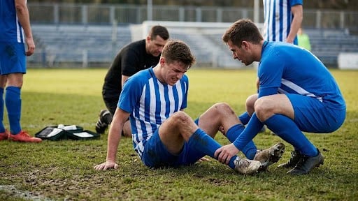
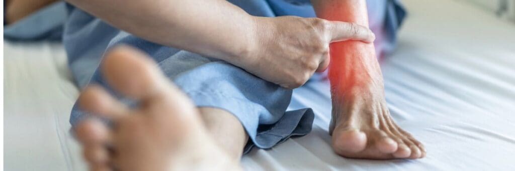
Osteochondral lesions are more common than many patients realize. Studies suggest that up to 50% of significant ankle sprains involve some degree of cartilage damage, yet clinicians often miss the lesion on initial assessment. In fact, the average time between injury and correct diagnosis can stretch to well over a year, simply because the symptoms so closely overlap with a routine sprain – swelling, tenderness, and a nagging ache that just will not go away. Because the symptoms of a talar dome lesion can closely mimic a routine ankle sprain, many patients spend months, or even years, being treated for the wrong condition before receiving an accurate diagnosis. If your ankle pain has not resolved after what was thought to be a straightforward sprain, an osteochondral lesion should be considered.
In younger patients and adolescents, a related condition called osteochondritis dissecans (OCD) can develop, in which a segment of bone and cartilage gradually separates from the talar dome and sometimes breaks free into the joint as a loose body. This is distinct from a traumatic osteochondral lesion in an adult, where a single forceful event – such as a severe ankle roll – is typically the trigger. Understanding which type of lesion you have is the first step toward choosing the right treatment path, and it is exactly the kind of nuanced assessment Dr. Bob Baravarian and his team are known for. Identifying this variant early matters, as timely treatment in adolescents can often preserve the native cartilage entirely.
Who is at risk? Causes and risk factors
The most common cause of an osteochondral lesion is a traumatic ankle injury, particularly an ankle sprain or fracture in which the bones of the ankle joint are compressed or twisted against one another. The impact can shear or crack the cartilage surface and bruise the bone beneath it.
However, not every osteochondral lesion follows a single dramatic injury. Repetitive microtrauma, such as the cumulative loading experienced by distance runners, basketball players, and dancers, can gradually degrade the cartilage-bone interface. Other contributing causes include:
Avascular necrosis, where a disrupted blood supply causes bone tissue to die, weakening the subchondral layer beneath the cartilage.
Chronic ankle instability, which allows abnormal joint motion that repeatedly stresses the same area of the talar dome.
Repetitive motion and cumulative stress on the joint from high-impact sport or occupational loading.
Certain groups face a meaningfully higher risk of developing an osteochondral lesion:
Athletes in high-impact sports (football, basketball, soccer, gymnastics, and distance running) who sustain frequent ankle sprains or land heavily on the joint.
Individuals with chronic ankle instability, where ligament laxity allows the talus to shift abnormally within the ankle mortise.
Adolescents and young adults, whose developing bone may be more susceptible to osteochondritis dissecans.
People with a history of prior ankle sprains that were not fully rehabilitated, leaving residual instability.
Those carrying excess body weight, which increases compressive forces across the ankle joint with every step.
Lesion location also matters clinically. Medial lesions (on the inner side of the talar dome) tend to be deeper and cup-shaped, often associated with repetitive stress, and are more likely to develop subchondral cysts. Lateral lesions (on the outer side) are more commonly linked to a single traumatic event and tend to be shallower and wafer-shaped. Understanding which side is affected helps guide both the surgical approach and the expected recovery.
Symptoms of a talar dome lesion
One of the reasons osteochondral lesions are so frequently misdiagnosed is that their symptoms often appear to resolve after the initial injury, only to return weeks or months later once the patient resumes normal activity. This pattern of apparent recovery followed by persistent or worsening pain is a key warning sign.
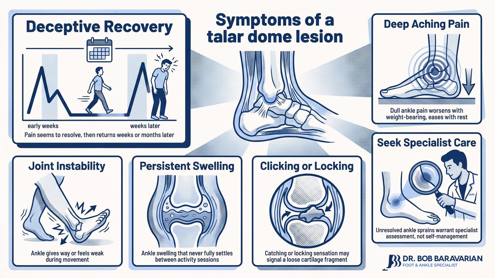
Patients with talar dome lesions may experience:
Pain that resolves after the initial trauma but then returns with activity.
Deep, aching ankle pain that worsens with weight-bearing and improves with rest.
Swelling around the ankle joint that does not fully settle between activity sessions.
Ankle instability, giving way, or a sense of weakness in the joint.
A clicking, catching, or locking sensation within the ankle may indicate a loose cartilage fragment.
If you recognize this pattern, particularly if you have a history of ankle sprains that never quite healed, seek a specialist assessment rather than continuing to manage what may be an incorrectly diagnosed sprain.
Diagnosing an osteochondral lesion of the talus
Accurate diagnosis begins with a thorough clinical history and physical examination. A specialist will ask about the mechanism of injury, the timeline of symptoms, any prior ankle sprains or fractures, and how the pain behaves with different activities. The physical examination assesses the range of motion, joint-line tenderness, and signs of instability.
Imaging is essential to confirm the diagnosis and determine the extent of the lesion:
X-ray can identify bony fragments or advanced lesions, but will miss early-stage or purely cartilaginous damage.
MRI is the gold-standard imaging tool for osteochondral lesions. It reveals the cartilage surface, subchondral bone integrity, bone marrow edema, and any cystic changes beneath the lesion. MRI findings are commonly graded using the Hepple MRI classification (Stages 1-5), ranging from cartilage signal change only (Stage 1) to a displaced fragment with a cystic lesion of the talus (Stage 5).
CT scan provides superior detail of the bony architecture and is particularly useful for measuring lesion size and planning surgery.
Lesion size is one of the most important factors in determining treatment. As a general guide, lesions smaller than approximately 1.5 cm² are often amenable to bone marrow stimulation techniques, while larger osteochondral defects typically require cartilage transplantation or grafting procedures. If you have an MRI report and are unsure what the findings mean for your treatment options, a specialist consultation can translate them into a clear, personalized plan.
Osteochondral lesion grading and MRI classification
So you’ve had an MRI, and your report mentions something like “Stage III osteochondral lesion” – what does that actually mean? Once imaging confirms an osteochondral lesion, your doctor will use a grading system to understand how deep the damage goes and whether the cartilage fragment is still attached or floating free. The most widely used MRI-based system is the Hepple classification, and it gives your care team a much clearer picture than X-rays alone ever could.
If you’re curious about what an open MRI for the foot and ankle involves, it’s a straightforward, non-invasive scan – nothing to worry about.
Here’s how the Hepple stages break down in plain language:
Hepple Stage | What’s happening |
|---|---|
Stage I | Cartilage injury only – the bone beneath looks normal on MRI |
Stage II | Cartilage injury with underlying bone damage and swelling (bone marrow edema) |
Stage III | Detached fragment that hasn’t yet displaced from its bed |
Stage IV | Fragment fully detached and displaced – a loose body in the joint |
Stage V | Subchondral cyst formation beneath the cartilage surface |
Beyond the stage, lesion size matters enormously for treatment planning. Lesions smaller than 1.5 cm² often respond well to non-surgical care or minimally invasive procedures. Larger lesions – particularly those exceeding 1.5 cm² – are more likely to need a cartilage transplant procedure to restore the joint surface properly. This is exactly why a detailed MRI read by an experienced foot and ankle specialist, rather than a general radiologist, can genuinely change the treatment path you’re offered.
Staging: the Berndt and Harty classification
The Berndt and Harty classification system is the most widely used framework for staging osteochondral lesions of the talus. It describes the degree to which the cartilage and underlying bone have been disrupted, and it directly informs treatment decisions:
Stage I: The subchondral bone is compressed, but the cartilage surface remains intact. The lesion is stable and may not be visible on plain X-ray. MRI typically shows bone marrow edema. Non-surgical management is usually appropriate at this stage.
Stage II: A partial fracture of the subchondral bone has occurred, creating an incompletely detached fragment. The overlying cartilage may be damaged, but the fragment has not displaced. Non-surgical treatment is still attempted first, though surgical intervention may be needed if symptoms persist.
Stage III: The osteochondral fragment is completely detached from the underlying bone but remains in its original position within the crater. Surgical treatment is generally recommended, as the fragment is unlikely to heal without intervention.
Stage IV: The fragment is completely detached and has displaced into the joint as a loose body. This stage almost always requires surgery to remove or reattach the fragment and address the resulting defect in the talar dome.
Understanding your stage helps set realistic expectations. Stage I and II lesions treated conservatively have good outcomes in many patients, while Stage III and IV lesions, particularly those with large defects or subchondral cysts, benefit most from the advanced surgical and regenerative techniques described below.
Non-surgical treatments for talar dome lesions
For mild to moderate lesions, particularly Stage I and II injuries without displaced fragments, a structured non-surgical program is the appropriate first step. Non-surgical management is typically pursued for at least three to six months before surgery is considered, provided the patient is progressing and no loose bodies cause mechanical symptoms. If pain persists beyond this window, or if imaging shows the lesion is failing to consolidate, surgical intervention becomes the more appropriate path – but reaching that decision takes time, patience, and close monitoring.
Non-surgical options include:
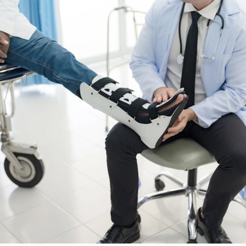
Immobilization with a non-weight-bearing boot or cast to offload the damaged area and allow the subchondral bone to stabilize.
Physical therapy to restore range of motion, strengthen the muscles supporting the ankle, and address any underlying instability once the acute phase has settled.
Nonsteroidal anti-inflammatory drugs (NSAIDs) to manage pain and reduce inflammation during the healing period.
Platelet-rich plasma (PRP) injections: PRP is prepared by concentrating the patient’s own blood platelets, which are rich in growth factors that stimulate tissue repair. When injected into the ankle joint, PRP creates a pro-healing environment that can support cartilage matrix regeneration and reduce inflammation. Because cartilage has a very limited natural blood supply, it heals poorly on its own. PRP compensates by delivering a concentrated burst of healing signals directly to the lesion site, essentially jump-starting a repair process the body struggles to initiate on its own. It is particularly useful as an adjunct to other treatments or in patients who are not yet surgical candidates.
Amniotic stem cell injections: Unlike PRP, which relies on growth factors alone, amniotic-derived preparations contain a broader range of regenerative signals, including cytokines and extracellular matrix components, that can modulate the inflammatory response and support tissue remodeling. These preparations also introduce scaffolding proteins that help organize new tissue growth, giving the repair process both a biological signal and a structural framework to build upon – something PRP alone cannot provide. These injections suit patients with early-stage lesions or those seeking to delay or avoid surgery.
Shockwave (Softwave) therapy: This non-invasive treatment delivers acoustic energy pulses to the damaged tissue, stimulating the body’s own repair mechanisms, improving local blood flow, and promoting the release of growth factors. It is a valuable option for patients who have not responded fully to rest and physical therapy alone.
Surgical treatment of talar dome lesions
When non-surgical management has not provided adequate relief, or when the lesion is large, unstable, or involves displaced fragments, surgery offers highly effective solutions. The choice of procedure depends on lesion size, stage, location, and the condition of the surrounding cartilage and bone. These include:
Berndt and Harty staging: what your stage means for treatment
Understanding where your osteochondral lesion falls on the Berndt and Harty classification scale is one of the most important steps in planning the right surgical approach. This four-stage system describes how far the lesion has progressed, from a subtle compression injury to a completely detached fragment floating inside the joint.
Stage I involves a small area of subchondral bone compression with the overlying cartilage still intact. The bone has been injured, but nothing has shifted. At this stage, conservative care – rest, immobilization, and regenerative therapies like PRP or Softwave – is almost always the first path forward.
Stage II describes a partially detached osteochondral fragment. The cartilage and underlying bone have begun to separate but remain connected. Non-surgical treatment is still attempted, though arthroscopic debridement becomes a realistic option if symptoms persist.
Stage III means the fragment is fully detached but has not yet migrated from its original position. At this stage, surgery is typically recommended. Arthroscopic debridement with microfracture works well for smaller Stage III lesions, while larger defects may call for an osteochondral autograft (OATS) procedure.
Stage IV is the most advanced presentation – the fragment has completely separated and is displaced within the joint. Surgical intervention is nearly always necessary, and depending on lesion size and bone quality, options range from OATS to fresh allograft cartilage transplant. Patients with Stage IV lesions can take real reassurance that, even at this stage, proven surgical solutions exist, and outcomes in experienced hands are consistently strong.
Arthroscopic debridement and bone marrow stimulation (microfracture)
For smaller lesions (generally under 1.5 cm²), arthroscopic debridement combined with bone marrow stimulation is the most common first-line surgical approach. During arthroscopic debridement, the joint is cleaned of loose debris, and any impinging tissue is addressed through small keyhole incisions. The surgeon then creates controlled microfractures in the exposed subchondral bone, allowing blood and bone marrow stem cells to flood the defect. The resulting clot matures into fibrocartilage, a repair tissue that fills the defect and restores a functional joint surface.
Fibrocartilage, while durable and clinically effective, is not identical to the original hyaline cartilage that covers a healthy talar dome. Hyaline cartilage has a more organized collagen structure and superior load-bearing properties. For this reason, microfracture is most appropriate for smaller defects in patients with good bone quality, and outcomes are generally excellent in this group, with studies reporting satisfactory results in 70-85% of patients at medium-term follow-up. It maps most naturally to Stage II and smaller Stage III lesions where the bone bed is healthy enough to support the repair process.
Osteochondral autograft transfer system (OATS)
For larger or cystic lesions where microfracture is unlikely to produce a durable result, the osteochondral autograft transfer system (OATS) offers a more robust solution. Healthy plugs of bone and hyaline cartilage are harvested from a low-load-bearing area of the patient’s own knee and transplanted into the talar defect. Because the graft comes from the patient’s own body, rejection risk is virtually eliminated, and the transplanted hyaline cartilage integrates well with surrounding tissue. OATS is particularly well-suited to larger Stage III and Stage IV lesions and to patients who have failed a prior microfracture procedure.
Allograft cartilage transplant
When the defect is too large for an autograft or harvesting from the patient’s own knee is not appropriate, an allograft cartilage transplant using donor tissue from a tissue bank is an excellent alternative. Fresh allograft tissue preserves viable chondrocytes (cartilage cells) and provides both the structural bone support and the hyaline cartilage surface needed to restore the talar dome. This approach is most commonly indicated for Stage IV lesions with large or cystic defects, and for patients presenting with a failed prior surgical repair – sometimes called revision surgery – where the original bone bed has been compromised. This approach is reserved for the most complex cases and delivers strong outcomes in experienced hands.
BioCartilage transplant for osteochondral lesion treatment
BioCartilage is one of the most exciting advances in cartilage restoration available today, and it represents a genuinely differentiated option for patients with osteochondral lesions of the talus. Developed from allograft cartilage extracellular matrix, BioCartilage is a micronized cartilage scaffold that is mixed with the patient’s own platelet-rich plasma (PRP) and applied directly into the prepared lesion site during an arthroscopic procedure.
The scaffold provides a three-dimensional matrix that mimics the natural environment of healthy cartilage. This matrix supports the migration and differentiation of the patient’s own repair cells into the defect, while the PRP component delivers a concentrated dose of growth factors to speed healing. The result is a biologically active repair tissue that more closely resembles native hyaline cartilage than the fibrocartilage produced by microfracture alone.
How the procedure works: After arthroscopic debridement of the lesion, the subchondral bone is prepared using microfracture to stimulate the release of bone marrow stem cells. The BioCartilage-PRP mixture is then applied to fill the defect precisely, conforming to the crater’s shape. A fibrin sealant secures the graft in place. The entire procedure is performed arthroscopically, allowing smaller incisions, less surgical trauma, and a faster return to activity than open grafting techniques.
Ideal candidates for BioCartilage are patients with contained osteochondral defects, typically in the small- to medium-size range, who have not responded to conservative management and who want to avoid the donor-site morbidity associated with autograft harvesting. It is also an excellent option for patients who have had a prior microfracture that has not produced a satisfactory result, as BioCartilage can be used to augment or revise the original repair.
Expected outcomes: Clinical studies and real-world experience with BioCartilage have shown encouraging results, with patients reporting meaningful pain reductions and improved function. Because the repair tissue produced is of higher quality than standard fibrocartilage, the durability of the result is expected to be superior over the long term. Patients typically return to low-impact activity within three to four months and to sport or high-demand activity within six to nine months, depending on lesion size and individual healing. Even patients with larger or more complex osteochondral lesions should feel reassured – when BioCartilage is combined with regenerative therapies like PRP or stem cell therapy, Dr. Bob tailors a treatment plan designed to give every patient the best possible path to lasting recovery. Read more about BioCartilage and whether it may be right for you.
What to expect after talar dome osteochondral lesion surgery
Because the talus has a limited intrinsic blood supply, healing takes longer than with most other bones. Patients should plan for a recovery measured in months rather than weeks, and following the rehabilitation protocol precisely is the single most important thing you can do to protect your result. The good news is that even large or complex osteochondral lesions have well-established, proven recovery pathways – and with personalized care and a tailored treatment plan, the vast majority of patients return to the activities they love. Here is your recovery timeline:
Weeks 0-2: Protection and pain management
Immediately after surgery, you will place the ankle in a splint or non-weight-bearing boot. You will use crutches and keep all weight off the foot. The priority during this phase is controlling swelling and pain through elevation, ice, and prescribed medication. Gentle range-of-motion exercises may begin within the first few days if your surgeon approves, to prevent stiffness without stressing the repair. Key milestone: swelling is visibly reducing, and pain is manageable with oral medication alone by the end of week two.
Weeks 2-6: Continued non-weight-bearing and early mobility
Weight-bearing restrictions continue during this phase for most procedures, particularly microfracture and BioCartilage, to allow the repair tissue to mature without compressive loading. Physical therapy begins in earnest, focusing on ankle range of motion, gentle strengthening of the calf and foot muscles, and maintaining cardiovascular fitness through upper-body or pool-based exercise where appropriate. Key milestone: achieving roughly 80% of normal ankle range of motion and tolerating light resistance band exercises without increased pain or swelling.
Weeks 6-12: Gradual weight-bearing and progressive strengthening
Controlled weight-bearing in a walking boot typically begins around six weeks, progressing to full weight-bearing in a supportive shoe as comfort and strength allow. Physical therapy advances to proprioception training, balance work, and progressive resistance exercises. Swelling should reduce steadily, and most patients notice meaningful pain improvement during this phase. Key milestone: walking comfortably in a standard shoe without assistive devices by week ten to twelve, with single-leg balance held for at least ten seconds.
Months 3-6: Functional rehabilitation
By three months, most patients walk comfortably and begin sport-specific movement patterns. Therapy focuses on dynamic stability, single-leg balance, and the controlled introduction of impact loading. Low-impact activities such as cycling and swimming are usually well tolerated. Running on flat surfaces may begin toward the end of this phase for patients who are progressing well. Key milestone: completing a straight-line jog for ten to fifteen minutes without pain or swelling flare-up, and passing a single-leg squat assessment with good control and no compensatory movement.
Months 6 and beyond: Return to sports
Full return to high-impact sport or physically demanding work is typically achieved between six and twelve months after surgery, depending on the procedure performed, lesion size, and individual healing. Arthroscopic debridement and microfracture patients often return to sport at the earlier end of this range, while those who have undergone BioCartilage augmentation, OATS, or allograft transplantation should expect a longer timeline to ensure the graft has fully integrated. Doing too much too soon remains the most common cause of setbacks, so milestone-based progression guided by your surgeon and physiotherapist is essential. Key milestone: passing a sport-specific functional test – such as a hop test or agility drill – with symmetrical performance on both limbs before being cleared for unrestricted return to play. Regenerative therapies like PRP or Softwave therapy may be incorporated during this phase to support tissue remodeling and help you cross the finish line faster.
Clinical outcomes: what the evidence shows
Patients researching osteochondral lesion treatment deserve honest, evidence-based information about what to expect. The published literature is encouraging across all major treatment approaches:
Arthroscopic debridement and microfracture produce satisfactory outcomes in approximately 70-85% of patients with small to medium lesions, with most reporting significant pain reduction and improved function at two to five years post-operatively.
OATS and autograft procedures show excellent results for larger lesions, with studies reporting good to excellent outcomes in over 85% of patients and high rates of return to sport at pre-injury levels.
BioCartilage augmentation is a newer technique, but early clinical data and surgeon-reported outcomes are consistently positive, with patients demonstrating superior cartilage fill quality on follow-up MRI compared with microfracture alone.
- Return to sports. Most athletic patients can return to sport after surgical treatment of osteochondral lesions, with published return-to-sport rates of 75-90% across procedures when rehabilitation is fully completed.
Even large or complex lesions, including those with subchondral cysts or prior failed treatment, have proven treatment paths. The key is matching the right procedure to the right patient, which is precisely where specialist expertise makes the greatest difference.
Why patients choose Dr. Bob Baravarian for osteochondral lesion treatment in Los Angeles
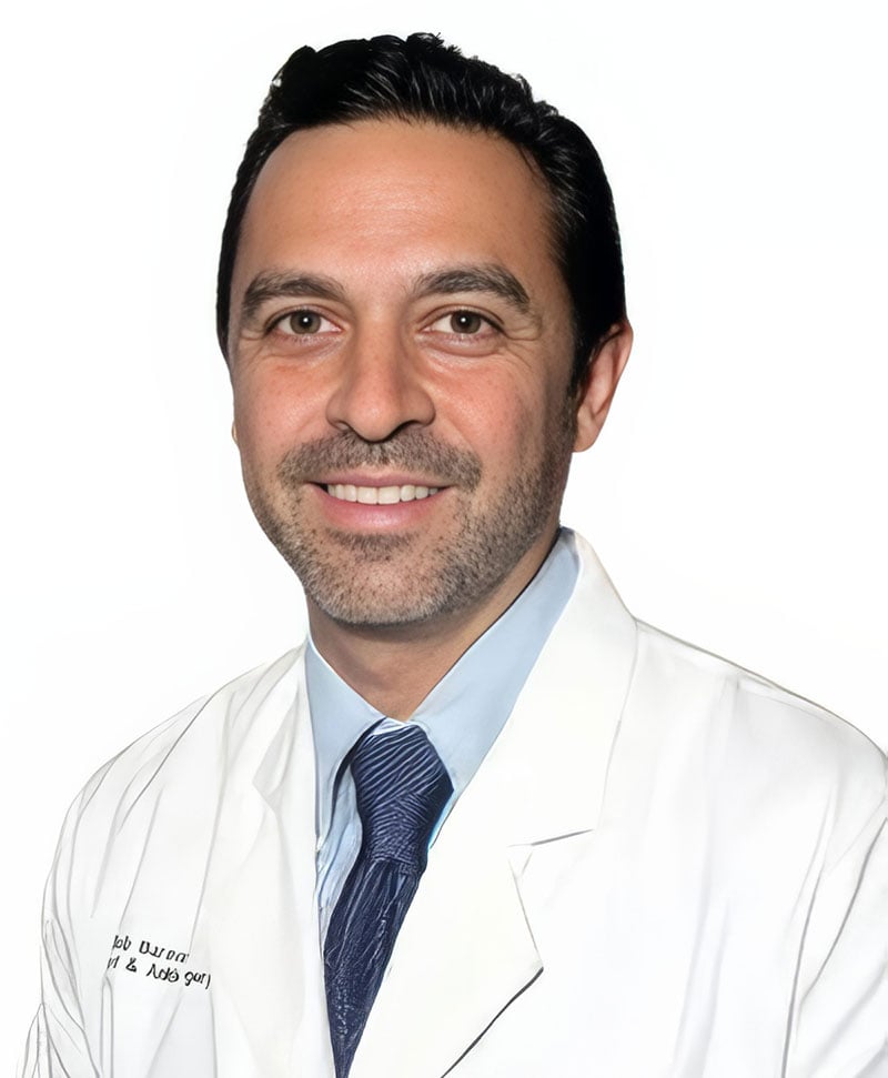
With over 25 years of experience, Dr. Bob Baravarian is nationally recognized as the surgeon other doctors trust for their toughest cases. He has dedicated his career to pioneering non-metal, minimally invasive, and regenerative techniques that deliver faster healing, less pain, and outstanding results, and his expertise in osteochondral lesions of the talus is among the most advanced available anywhere in the United States.
Dr. Bob takes the time to get to know every patient personally, tailoring each treatment plan as if he were caring for his family. Whether your lesion is a Stage I injury that may respond to regenerative injection therapy, or a complex Stage IV defect requiring BioCartilage augmentation or allograft transplantation, Dr. Bob will walk you through every option with clarity and confidence, so you can make an informed decision about your care.
His clinics are equipped with on-site X-ray, CT, and MRI imaging, allowing for same-visit diagnosis and treatment planning. He uses the full spectrum of treatments described on this page, from Softwave therapy and PRP injections to advanced arthroscopic cartilage restoration procedures, ensuring every patient receives the most appropriate, least invasive solution for their specific lesion.
Dr. Bob Baravarian is conveniently located throughout Southern California and the Los Angeles area, with clinics serving patients in and near Santa Monica, Sherman Oaks, Beverly Hills, West Los Angeles, the San Fernando Valley, El Segundo, the South Bay, LAX, Calabasas, Agoura Hills, and Downtown Los Angeles. New patients and those concerned about persistent ankle pain are warmly encouraged to call or schedule a consultation at (855) 557-5400.
Osteochondral Lesion FAQs
What is a talar dome fracture?

A talar dome fracture is sometimes used interchangeably with the term talar dome lesion. However, a lesion refers to the tearing or fracturing of the talar dome. In contrast, a talar dome fracture generally describes a condition in which a piece of cartilage breaks off from the talar dome.
Does a talar dome lesion need surgery?
If a talar dome injury is detected early enough, non-surgical treatment may relieve symptoms and support healing. However, surgery for talar dome lesions is often necessary if cartilage pieces have broken off and migrated into the joint, or if conservative treatment has failed to manage symptoms.
Will a talar dome lesion go away on its own?
Typically, talar dome injuries will not resolve on their own without intervention. Immobilization and rest may allow minor cases to heal. Meanwhile, other talar dome lesions may require surgical cleaning, microfracturing to promote healing, or bone grafting.