Professional dancers place a large amount of stress on their bodies, especially their lower extremities. In particular, ballet dancers are prone to developing foot and ankle problems.
Dancing en pointe or demi-pointe is a particular technique that many female ballet dancers perform and it can cause foot and ankle injuries. The ability to dance en pointe requires the dancer to undergo many years of training to build up her physical strength, balance and coordination. The technique involves excessive plantarflexion of the ankle and often puts the flexor hallucis longus (FHL) under a lot of strain, leading to tenosynovitis of the FHL tendon.
Tenosynovitis of the FHL is an entity that rarely occurs in the general population but can affect ballet dancers due to the strain on the tendon while the dancer is en pointe and demi-pointe. There are many factors that can predispose the patient to putting an increased amount of stress on the FHL and causing subsequent injury. These factors include ligamentous laxity, poor en pointe technique and pronation of the foot.
The FHL passes posterior to the medial malleolus deep to the flexor retinaculum along with the posterior tibial tendon, the flexor digitorum longus tendon, the tibial nerve and posterior tibial artery. The FHL then courses through a groove between the medial and lateral tubercles of the posterior portion of the talus. The tendon then passes under a fibro-osseous tunnel plantar to the sustentaculum tali before it courses toward its insertion into the plantar aspect of the hallux.
How is FHL tenosynovitis diagnosed through physical examination?
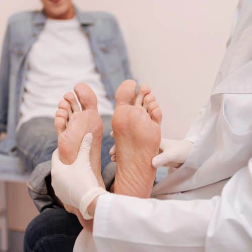
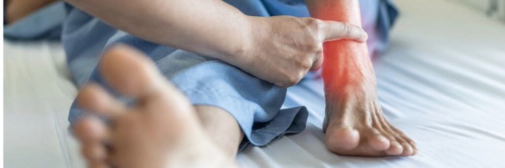
FHL tenosynovitis is diagnosed through physical examination by observing pain and swelling at the posteromedial ankle, exacerbated by jumping or going en pointe. Key findings include tenderness over the FHL tendon, pain with resistance to hallux flexion, and occasional snapping during passive range of motion. Patients often report a 'sticking' feeling in the posterior ankle. Care is taken to differentiate from os trigonum pain or tarsal tunnel symptoms. With longstanding cases, stenosis may cause the great toe to freeze in a plantar position, requiring manual release.
What imaging techniques are essential for diagnosing FHL tenosynovitis?
Essential imaging techniques for diagnosing FHL tenosynovitis include radiographs, MRI, and dynamic ultrasound. Radiographs rule out os trigonum, an accessory ossicle with similar symptoms. MRI is useful for evaluating FHL tendon tears, impingement from a low-lying muscle belly or accessory muscle, and further assessing os trigonum. Dynamic ultrasound is often the best diagnostic option for stenosis or impingement of a low-lying muscle belly, allowing real-time testing of the FHL tendon during range of motion. Combining ultrasound with MRI can help check for os trigonum problems or posterior ankle impingement.
What are the effective treatment options for FHL tenosynovitis?
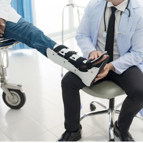
Effective treatment options for FHL tenosynovitis include conservative care and surgical intervention. Conservative treatment involves resting the area, reducing inflammation with ice and NSAIDs, physical therapy (stretching, strengthening, massage, ultrasound), and immobilization with a cast or boot. Corticosteroid injections are rarely performed due to rupture risk. Surgical treatment is indicated when conservative therapy fails, consisting of releasing the FHL tendon retinaculum, debriding synovitic tissue, repairing tears, and debriding low-lying muscle belly or accessory flexor digitorum longus muscle belly to reduce impingement.
Rarely should one perform injections of corticosteroids secondary to the risk of weakening or rupturing the tendon. If you are performing a corticosteroid injection in the sheath of the FHL, be sure to immobilize the patient in order to reduce the risk of rupturing the tendon.
Surgical treatment is indicated when symptoms persist despite conservative therapy. Surgery consists of releasing the FHL tendon retinaculum posterior to the medial malleolus and continuing down to the level of the sustentaculum tali. Put the great toe through range of motion in order to visualize unrestricted motion of the tendon.
Inspect the FHL at the time of surgery. Debride any synovitic tissue and repair any longitudinal tears of the FHL. If there is a low-lying muscle belly of the FHL, one can also debride this to reduce impingement while the ankle is in a plantarflexed position.
There is a possibility of an accessory flexor digitorum longus muscle belly, which may be impinging on the FHL. One can debride this muscle at the time of surgery to prevent further impingement on the FHL. The complications of the procedure include stiffness and recurrence of the tenosynovitis with an inadequate release as well as possible subluxation of the FHL.
What are the key takeaways for FHL tenosynovitis in dancers and athletes?
The key takeaways for FHL tenosynovitis in dancers and athletes are that posterior ankle pain, often overlooked, frequently stems from the FHL tendon. It is crucial to consider FHL issues in the differential diagnosis for athletes, especially dancers. With accurate examination and diagnostic testing, both conservative and surgical care, followed by postoperative protection, consistently lead to excellent outcomes. This article was contributed by Dr. Baravarian, Assistant Clinical Professor at UCLA School of Medicine and Chief of Podiatric Foot and Ankle Surgery at Santa Monica UCLA Medical Center, and Dr. Yau, a Fellow at the University Foot and Ankle Institute in Los Angeles.
Dr. Baravarian is an Assistant Clinical Professor at the UCLA School of Medicine. He is the Chief of Podiatric Foot and Ankle Surgery at the Santa Monica UCLA Medical Center and Orthopedic Hospital, and is the Director of the University Foot and Ankle Institute in Los Angeles.
Dr. Yau is a Fellow at the University Foot and Ankle Institute in Los Angeles.