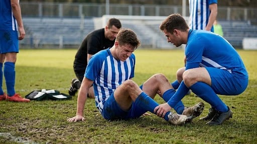
Osteochondral fractures of the talus have been a challenging and often poorly treated problem in podiatry practices. They are often undiagnosed at the initial time of injury and will cause pain with increased activity. This subsequently leads to patients presenting with an ill-defined ankle pain that can be challenging to diagnose.The current thinking and treatment regimen for osteochondral lesions has changed dramatically from a decade ago and several excellent options are available to facilitate improved outcomes. Accordingly, let us take a closer look at the possible causes of osteochondral lesions of the talus, diagnosis and current treatment options.Osteochondral lesions are most often traumatic in nature. The most common underlying cause of injury is an ankle sprain with rotation of the talus causing a jamming of the dorsal talar articular surface against the underlying distal surface of the tibia or fibular. This rotation/shear force may cause a destruction of the overlying cartilage with or without underlying fracture of the talar bone.
In general, medial lesions are more posterior and deeper while lateral lesions are more superficial and anterior. The reasoning is quite simple. Posterior medial lesions result from a plantarflexed and internally rotated ankle position whereas lateral lesions result from a dorsiflexed and externally rotated ankle position.When patients present with an ankle sprain, radiographs of the ankle are essential as they will help diagnose large osteochondral fractures or loose bodies in the ankle, which may be the result of an osteochondral fracture. However, in most cases, osteochondral lesions are not visible on standard radiographs and do not receive treatment until follow-up visits with the patient having continued, non-resolving ankle pain.For certain patients, such as those involved in a car accident with jamming of the ankle on the brake pedal, the underlying injury may be the result of some other form of trauma to the talus.Indeed, patients will often present at three months to several years after an initial underlying injury with chronic pain and swelling of the ankle. The patient history will often refer to clicking or locking of the ankle, and sharp pain with certain rotation motions. Patients will often relate that they have less pain in a restricting ankle brace and feel more stable.With medial lesions, there is pain along what seems to be the posterior tibial tendon or medial malleolar region while lateral lesions present with anterior lateral ankle gutter pain similar to what occurs with impingement lesions or synovitis lesions of the lateral gutter.
What You Should Know About Diagnostic Imaging
When it comes to diagnosing osteochondral lesions, it is best to obtain a magnetic resonance image (MRI) of the ankle. There is debate on the use of dye in the MRI. I prefer not to use dye on the standard MRI. The rationale behind using dye is that it helps pick up faint cartilage lesions or loose lesions that are well seated by either lining the damaged cartilage with a dye layer or by soaking under the lesion. These are very subtle cases and often still difficult to pick up with dye injection. In most cases, physicians utilize a standard MRI to check for ankle pathology.With this patient population, magnetic resonance imaging findings often show damage to the underlying cartilage and possibly the bone. Cyst formation may occur with long-standing lesions that project into the talus.If the MRI is not clear and you suspect superficial cartilage damage, a repeat MRI with dye injection may be a suggestion. In most patients, MRI can over-read the size and depth of the lesion due to the marrow edema associated with the trauma. Therefore, it is essential to look at the actual MRI films to pick up the size of the actual damage and not the surrounding marrow changes.If there is a concern that the lesion may be much larger or, more often, much smaller than the MRI may show due to edema, you can also order a CT scan to check the actual size of the osteochondral lesion. Determining the true underlying size of the osteochondral lesion is essential to treatment.
What are the treatment options for osteochondral lesions?
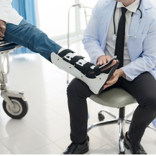
Treatment options for osteochondral lesions vary widely depending on the lesion's size, depth, and whether it's painful. Not all osteochondral lesions require treatment; some incidental, non-painful findings may be left alone. For those that do, options range from conservative measures like activity modification, custom ankle-foot orthotics, and viscosupplementation, to surgical interventions such as arthroscopic debridement with subchondral drilling, cartilage cell transplantation, bone and cartilage transplantation (using autograft or allograft), and bone grafting for cystic lesions. The choice of treatment is highly dependent on the specific characteristics of the lesion, including its size, depth, and the presence of cystic damage or cartilage damage.
What are the key considerations for selecting osteochondral lesion treatment procedures?
In the case of a superficial lesion that is less than 0.5 cm in width with cartilage damage, debridement and drilling are usually successful and fairly simple. If the lesion is 0.5 cm to 1 cm and superficial with cartilage damage, one may opt for drilling or cartilage cell transplantation. Lesions that are over 1 cm and superficial require either cartilage cell transplantation or osteochondral autograft transplantation (OATS) of bone and cartilage plug. In these patients with cartilage damage and superficial lesions, there is no real cystic damage. Therefore, cyst treatment is not an issue.As the lesion deepens, treatment options vary. When there is bone and cartilage damage that is deeper than just the superficial cartilage, the best way to consider treatment is to consider the size of the lesion. Once a lesion consists of bone and cartilage damage, the width dictates treatment.Since the surface area of the talus is only 4 cm wide, lesions over 1 cm wide and through the subchondral bone do not do as well with debridement and drilling or cartilage cell grafting as more superficial lesions. If the lesion is less than 0.5 cm wide but fairly deep with cartilage damage, bone and cartilage grafting may be necessary. It is best to consider depth and width.One should also consider if the region may possibly fill with stable fibrocartilage via subchondral and bone drilling. Alternately, the surgeon may need to fill the region with graft. Deeper lesions require more aggressive treatment and cartilage cell transplants do not work well.Therefore, as the lesion gets deeper and larger, we tend to move further away from drilling and closer to bone and cartilage plug grafting.The only instance that is different is a cystic lesion with the superficial cartilage and bone being fairly stable and intact over the lesion. This type of lesion is rare. It is due to a small crack in the cartilage and bone that drains the joint fluid into the talus bone and causes a cyst.As the cartilage and bone are stable, one can perform packing of the cyst through the sinus tarsi through a plantar approach. The surgeon can drain and pack the cyst without damage to the superficial cartilage and bone.
What About Postoperative Rehabilitation?
Rehabilitation of osteochondral lesions is critical to proper outcome. When it comes to drilling, lay down fibrocartilage with constant passive motion. After two to three days of protection, the patient may begin rehabbing the ankle with a passive range of motion machine during the waking hours of the day. Although range of motion is allowed, one should ensure the patient is non-weightbearing for four to six weeks depending on the size of the lesion.Cartilage cell transplants require non-weightbearing for six weeks and no motion for the first four to six weeks in order to allow the cells to adhere. The patient may subsequently start gentle passive range of motion and protected weightbearing is critical for an additional two months.Cartilage and bone plug grafting require ingrowth of the bone and cartilage into the surrounding tissue. Usually a tibial or fibular osteotomy is also necessary to access the lesion and may dictate the weightbearing period. On average, six to 12 weeks of non-weightbearing is required for adequate healing but one may initiate passive range of motion as early as four weeks after surgery. Rigid fixation of the osteotomy access region will allow early range of motion.
What are the key conclusions regarding diagnosing and treating osteochondral lesions?
The key conclusions regarding diagnosing and treating osteochondral lesions emphasize that the size and depth of the lesion are essential to proper treatment, with superficial and smaller lesions generally having better outcomes than deeper and larger ones, which require more aggressive interventions. Additionally, aggressive and early range of motion is crucial for better cartilage growth and reduced scar formation. With proper diagnosis and tailored treatment, osteochondral lesions can heal effectively, leading to high patient satisfaction. Dr. Baravarian, an Assistant Clinical Professor at the UCLA School of Medicine and Chief of Foot and Ankle Surgery at Santa Monica UCLA Medical Center, provides expert care in this field.