Locations
Dr. Bavarian discusses Posterior Tibial Dysfunction
Posterior Tibial Dysfunction
The tibialis posterior tendon attaches the calf muscle to the bones inside the foot. It passes through the inside of the ankle before continuing up the lower leg. It supports the foot and arch while walking.
Flat feet or constant stress in the midfoot can cause the tendon to fray and tear, resulting in posterior tibial tendon dysfunction (PTTD). The condition is also the most common cause of adult-acquired flatfoot deformity, as the weakening of the tendon causes the arches to fall. About 80% of adult-acquire flatfoot cases are thought to be caused by PTTD.
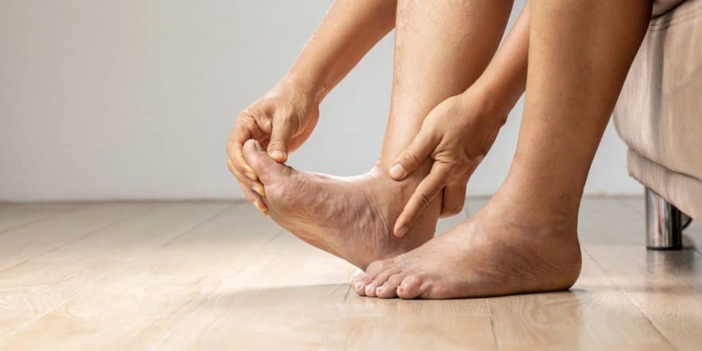
What are the posterior tibial tendon tear symptoms?
In its early stages, posterior tibial dysfunction can be characterized by an inflamed tendon. Because the tendon runs along the back of the ankle near the Achilles tendon, it can sometimes be mistaken for Achilles tendinitis.
In the later stages of PTTD, the injured tendon is unable to fully support the arch of the foot, and the condition progresses to posterior tibial tendon insufficiency. The arch falls because the tendon is no longer able to do its job.
Tendon degeneration, and PTTD in particular, is prevalent in people over 40 with flat feet and affects as many as five million Americans. It may also occur due to an acute injury, such as a fall or overuse (especially for those involved in high-impact sports). Other risk factors for PTTD include hypertension, diabetes, and obesity.
If you’re experiencing any of the following symptoms, get in touch with Dr. Baravarian to see if you’re suffering from PTTD:
- Swelling
- Pain around the hindfoot
- Ankle pain
- Pain that worsens during high-impact activities
- Foot pain that has moved to the outside of the ankle
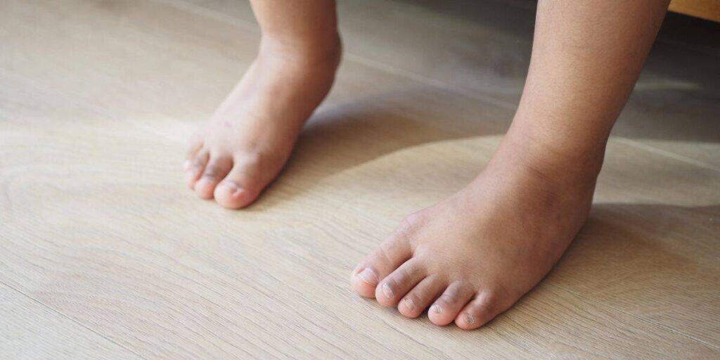
- Flattening of the arches
- An ankle joint that rolls inward when you walk
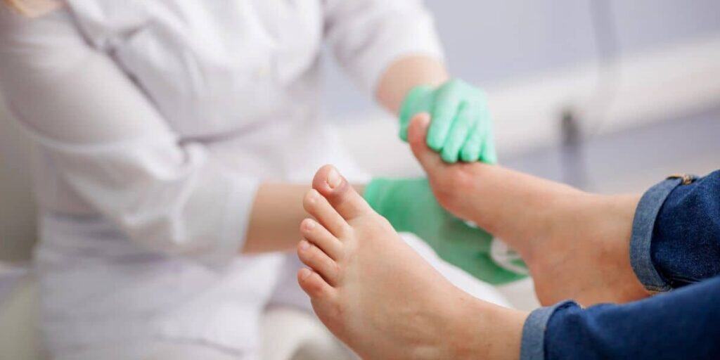
How does Dr. Baravarian diagnose a tibial tendon tear?
The first step in diagnosis is a review of your symptoms and medical history, along with a physical examination of your entire foot (forefoot, midfoot, and hindfoot). Dr. Baravarian will check for swelling along the posterior tibial tendon, changes to your foot shape, and how your feet and ankles move during weight-bearing activities. Specifically, we’ll see how your feet and ankles respond to simple heel raises
Dr. Baravarian also has access to X-rays, MRIs, CT scans, and ultrasounds to get a clear view of your bones and tendons, which can also help rule out other possible causes of your symptoms.
What is the posterior tibial tendon dysfunction treatment?
PTTD is a progressive condition that worsens with use and age. Therefore, Dr. Baravarian advises treating the condition as early as possible. The earlier PTTD is caught and treated, the easier and less invasive the treatment will be.
Non-surgical treatments for tibial tendon dysfunction
Stage I of PTTD is characterized by inflammation of the tendon, but without fraying or tearing. If the problem is detected at this stage, before the tendon tears, it can be alleviated with simple supportive therapies.
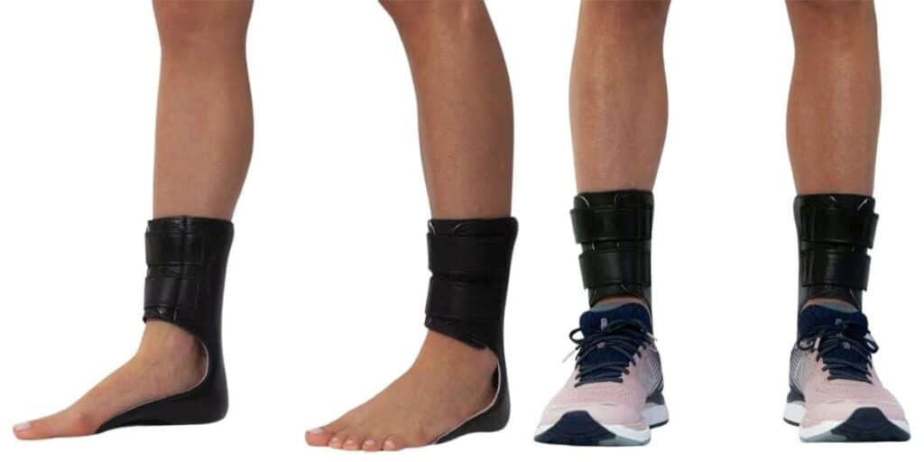
Orthotic devices, such as ankle braces, shoe inserts, splints, and custom orthotics, can support the tendon, improve your gait, and facilitate ankle healing. Combining these devices with physical therapy to strengthen your muscles and tendons, improve their flexibility, and prevent future injury.
Dr. Bob can also add conservative treatments for pain management. These treatments may include steroid injections, icing your foot, and non-steroidal anti-inflammatory drugs (NSAIDs) such as ibuprofen.
At the beginning of treatment, you may need to immobilize the joint with the use of a walking boot or an ankle-foot orthosis brace. Keeping the joint from moving can reduce inflammation in the soft tissues and ligaments.
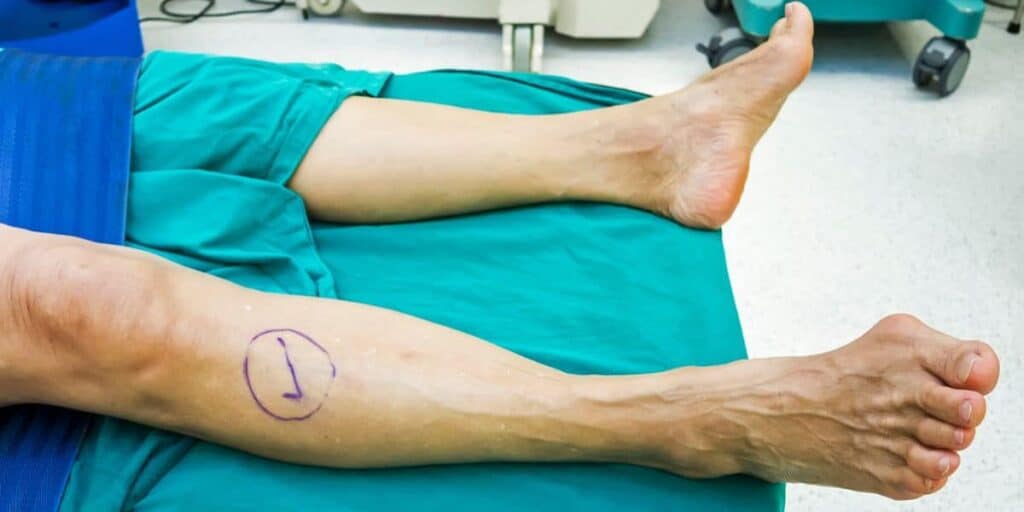
Posterior tibial tendon surgery
If the tendon tears, your PTTD condition is considered stage II or worse. Stage III and Stage IV include not only pain and changes in your gait but also foot deformity and degeneration of the ankle joint. A torn posterior tibial tendon generally requires PTTD surgery to restore the tendon and prevent further collapse of the arch.
Minimally invasive surgical options
Dr. Baravarian always starts with the least invasive treatment option possible. One such option is a tenosynovectomy. With this technique, the inflamed tissue is gently removed from around the tendon. Dr. Baravarian tends to use coblation Topaz therapy to accomplish this. This form of therapy is minimally invasive and uses radio frequencies to break up scar tissue around the damaged tendon. A tenosynovectomy alone can help reduce inflammation, but it won’t solve the issue of the tendon being disconnected.
If your tendon needs to be reattached to the main bone, keyhole surgery may be used to remove damaged parts of the tendon and then reattach the healthy portions using a specialized screw. The screw can be placed on the calcaneus (heel bone), and it can be adjusted to the tension of the tendon to restore your foot arch. In some cases, stem cells are injected into the surgical area to help strengthen the tendon and facilitate its fusion to the screw.
Another option for creating a new arch is an osteotomy, in which the bones of your heel and midfoot are reshaped to recreate the arch and relieve pain.
Surgical options for severe PTTD
When a tendon becomes too weak to support itself, it may require the extra support of another tendon. Using half of an adjacent tendon, a tendon transfer is done to recreate a stable posterior tibial tendon. Typically, another tendon in the foot will be transferred, such as the flexor digitorum longus (FDL) that moves the pinky toe or even the flexor hallucis longus (FHL) that moves the big toe.
However, there are times when a simple repair of the tendon is not enough to improve arch support. Flat foot reconstruction surgery can entail moving and reshaping bones. One instance is lateral column lengthening, in which a bone is taken from your hip and fastened to the outside of your heel bone to form an arch. Additionally, arthrodesis may be required if arthritis has set in. During this procedure, your doctor will reshape and realign the bones to achieve a proper foot arch. Once they are realigned, the cartilage between them is removed, and they are fused.
Dr. Bob will help determine the best treatment for you.
Why choose Dr. Baravarian for your tendon care?
Dr. Baravarian is active in sports, so he understands the stress that physical activity puts on the feet. He is a nationally recognized expert with decades of experience in foot and ankle tendon care, utilizing the latest technologies available. His goal is to help you get back on your feet, pain-free.
By offering a full spectrum of workup, conservative, surgical, and recovery options, Dr. Baravarian is truly taking care of you in a state-of-the-art manner, without the need to visit multiple locations.
To schedule a consultation, please call (855) 557-5400 or make an appointment now.
Dr. Bob Baravarian is conveniently located throughout Southern California and the Los Angeles area—at locations in or near Santa Monica, Sherman Oaks, Beverly Hills, West Los Angeles, the San Fernando Valley, El Segundo, the South Bay, LAX, Calabasas, Agoura Hills, and Downtown Los Angeles.