 Minimally invasive techniques in foot and ankle surgery have gained popularity over the years. Although an open incision system is often optimal, in the case of Achilles tendon tears, the best option truly is a minimally invasive approach.
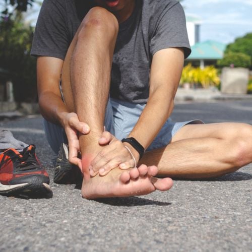
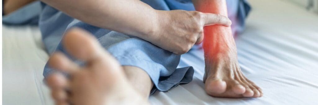
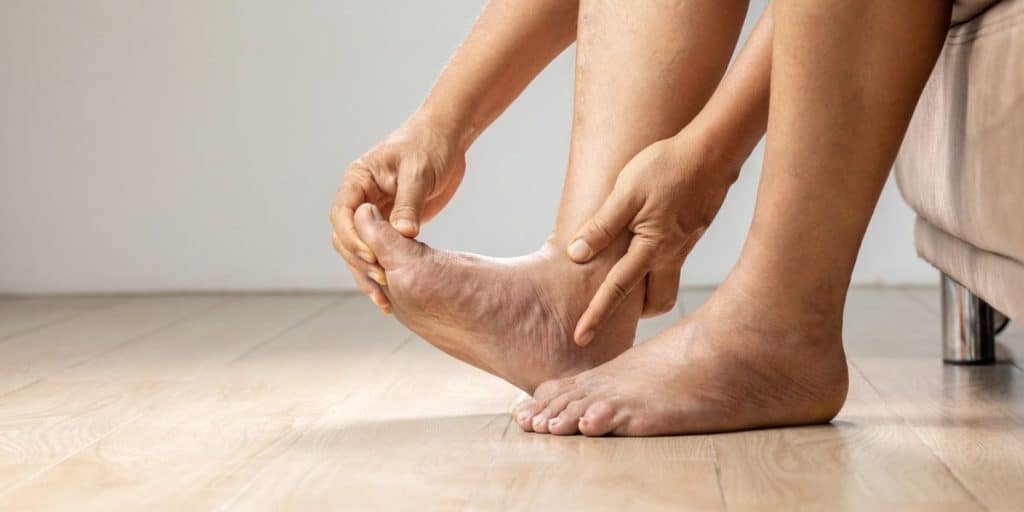
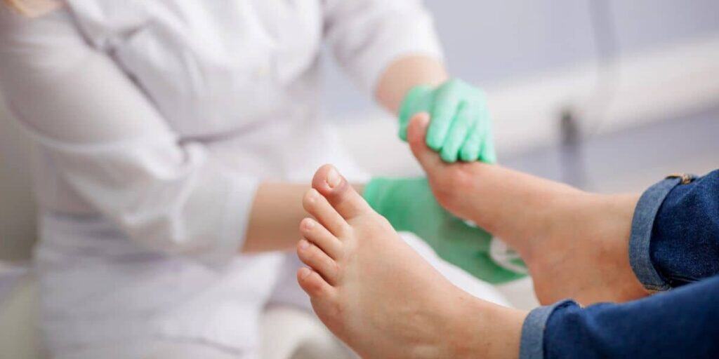
The Achilles tendon is the largest and strongest tendon in the body. A rupture of the tendon can occur with a sudden strain or stretch of the tendon, resulting in a loud “pop” and pain in the back of the ankle/heel. Patients will often note the feeling of being kicked or hit in the back of the leg. After a short period of time ranging from a few hours to a few days, the pain can resolve as the tendon is completely torn and does not assume any further strain, which makes some patients feel they did not injure themselves. What patients often note, however, is a feeling of weakness with push off and difficulty with single heel raise.
Repair of the Achilles tendon rupture is critical for proper function of the foot and ankle. It is rare not to repair a tear and the only cases of conservative care are often in poor surgical candidates and patients who have a sedentary lifestyle. In order to return to full activity, surgical repair of the tendon is essential.
Recognizing The Advantages And Disadvantages Of Open Achilles Repair
For years, surgeons have repaired the Achilles tendon through a large open incision. This technique requires full exposure of the tendon and opening of the sheath around the tendon, which prevents scar formation. Furthermore, the surgeon performs the repair in the damaged area of the tendon, which is the weakest and most problematic region. Often, surgeons utilize a Krackow suture technique, which is a running locking suture that runs up one side of the tendon and down the contralateral side. One would repeat this on the opposite side of the tear and then repair the tendon through a tie stitch at the tear region.
This type of repair has been the best open option but it is hard to get the tendon to have proper strength, and also hard to have the suture and knot not slip as the tendon is somewhat friable and weak in this region.
Our experience with an open repair and Krackow stitching has been good but not excellent. We have seen “stretch elongation” of the repairs, which I believe is due to the suture material being in the severely damaged region of the tendon. We also see more scar formation and thickening of the tendon itself related to the size of the incision, and stripping of the peritenon, which is often difficult to repair fully. Our results have been good but not consistently excellent with the open repair and that is why we looked for a better option.
The Percutaneous Achilles Repair: A Better Option?
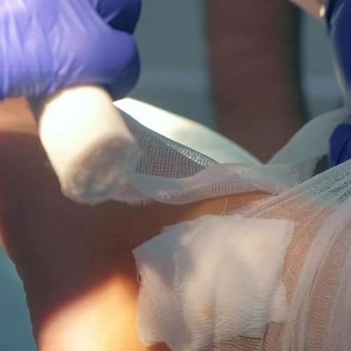
One can perform a percutaneous minimally invasive repair through a 1 cm incision. Place the stitches in the strong and healthy area of the tendon proximal and distal to the region of the tear. This allows grabbing of the healthy tendon and brings the tendon together in a stronger and more stable fashion. Then suture together the split ends of the tendon with small stitches locally in the 1 cm area for optimized repair.
There is an option of tying the knots in the tear area by passing the suture through the proximal and distal stumps. If the tear distally is close to the calcaneus and not enough tendon is present for suture and/or the surgeon prefers to avoid a local suture knot, place the proximal sutures into the calcaneus with a PushLock anchor system (Arthrex). This anchor system allows great tensioning and also removes the need for knots at the tear site.
Through the use of a 1 cm minimally invasive incision, we are able to avoid exposure of the tendon, which decreases scar formation and wound healing issues. We are also able to repair the tendon in the healthy region and not in the torn region by passing the stitches through the healthy tendon ends, and then bringing the tendon together. This allows a more rapid recovery, earlier physical therapy and a quicker return to activity.
In comparison to an open procedure, which usually requires physical therapy at about five to six weeks post-op in order not to overstrain the tendon, we have been able to get patients into physical therapy and out of a cast consistently at the three-week mark with the percutaneous repair technique.
Patients can bear weight at three weeks with a controlled ankle motion (CAM) walker boot in a plantarflexed position. We place 1/4 inch lifts in the boots stacked to a full inch at first and remove 1/4 inch per week, moving to flat at seven weeks. Patients remove the boot at eight to 10 weeks post-op depending on the amount of damage and quality of the tendon at the time of repair.
Anchoring the proximal repair distally to the calcaneus allows an even more rapid recovery as the suture is strongly attached to the calcaneus and one does not have to worry about knot slippage. I am concerned about potential over-tightening of the tendon as the suture material has little give with the distal anchoring and I have been watching this closely. However, I have not seen any adverse consequences with calcaneal anchoring to date.
A Closer Look At Our Achilles Treatment Protocol
Our current protocol for Achilles repair is to obtain consent from the patient for open as well as percutaneous repair. This allows us to take care of any problems that may occur with a minimally invasive technique, especially in a tear that is older with greater retraction. To date, I have performed over 30 percutaneous repairs in the past two years. I have only had to convert two repairs to an open procedure and both of these tears were over three months old. In both cases, I needed to free the tendon of scar tissue and it was difficult to do with a percutaneous incision.
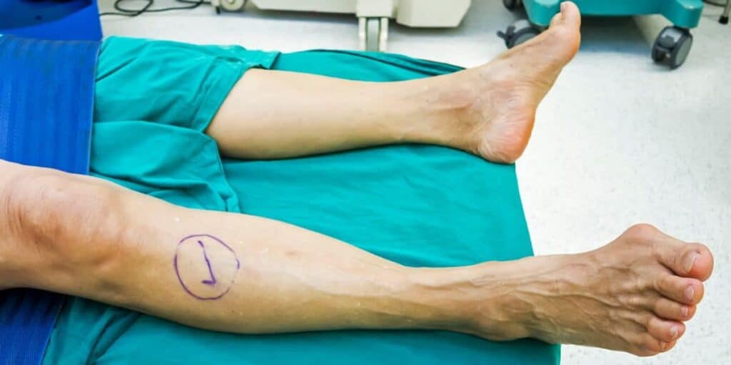
I will place an incision of approximately 1 to 2 cm longitudinally and slightly proximal to the tear region. The more proximal stump of tear is harder to identify and pull down than the distal stump. The incision needs to be large enough to fit an index finger. Open the peritenon only in the area of the incision and use an index finger to free up scar around the tendon. Use an Allis clamp to grasp the proximal tendon and pull it distally. Clean up the tendon end and remove sever-related damage to the tendon. Take care not to remove too much tendon at this time as one can do this later if necessary.
Place sutures in the proximal tendon in the region of healthy and non-torn tendon. Pull the sutures into the incision site and perform the same procedure to the distal stump. Then repair the tendon and suture the ends together. If the distal stump is poor quality or small in nature, we do not place sutures through the distal stump and instead anchor the proximal sutures into the calcaneus. Place the tendon in an almost maximally planatarflexed position and make it extra tight as it will lengthen with therapy and time. Take care to place the tendon slightly less tight if you are utilizing the calcaneal anchor technique.
Assessing The Results With Percutaneous Repair
To date, we have not had a single patient who has been unable to do a single heel raise at six months post-op. This is an amazing number. Our in-office patient questionnaire has resulted in over 95 percent of patients being happy with full return to activity at one year and no restriction in activity. We have performed the surgery on Division I college athletes several times with full return to activity.
The only complicating factor I have seen is irritation of the incision site by the suture knots on four patients. In all cases, the fiber knot was rubbing the skin incision raw, which resulted in a small blister. I easily opened the area without the need for anesthesia and cut and removed the knot in the office with full healing. This usually occurred as the patient progressed in activity at approximately three months post-repair.
In Conclusion
The percutaneous Achilles tendon repair system is the ideal approach to repair an acute Achilles tendon tear. It allows a very strong repair, limits exposure of the tendon, limits wound healing issues and scar tissue, allows a quicker return to physical therapy, and allows a more rapid return to full activity. In very rare cases, one may need to lengthen the incision due to severe damage to the tendon or severe retraction of the tendon ends, and this process is possible without an issue if the procedure needs to go from minimally invasive to open.
Our recovery process has often resulted in weightbearing and the initiation of physical therapy at three weeks post-op, and return to sports activity at about three months. This is approximately three months earlier than an open technique and is a significant improvement.
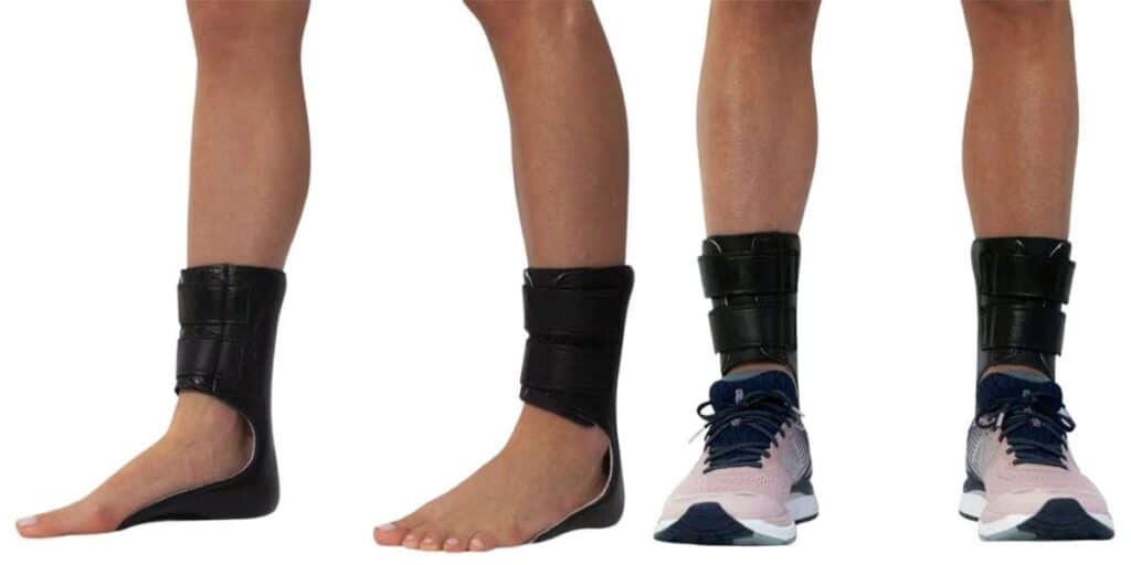
Furthermore, our rate of complications has been less than 1 percent with the minimally invasive technique and we are proud to note that 97 percent of our patients have returned to their previous level of activity with no long-term complications. The only patient who did not have a full return to activity was an older patient with a delayed repair who had some weakness of the tendon but was very happy overall with his outcome and ability to walk pain-free. His main issue was difficulty with paddle tennis, which was helped by an ankle foot orthotic brace to stabilize him and make him feel steadier.
I urge those repairing Achilles tendon tears to consider a minimally invasive technique. It is the best option we have found for patients and our outcomes have been outstanding.
Dr. Baravarian is an Assistant Clinical Professor at the UCLA School of Medicine, and the Director and Fellowship Director at the University Foot and Ankle Institute in Los Angeles