Patients with pediatric flatfoot deformity may present commonly to foot and ankle surgeons’ offices and the condition can prove fairly complex to work through. Although I personally find a majority of patients will improve and stabilize with the use of over-the-counter insoles or orthotics to alleviate their foot and ankle pain, there is a subset of pediatric flatfoot cases that require surgical intervention to address the deformity. In my experience, these patients have multiplanar deformities, which require correction through different procedures for different needs.
Although there are a multitude of procedures available, there are a select few that I have found to be my go-to options for pediatric flatfoot correction, and I will share them, along with relevant evaluation and examination pointers, in this article.
Considerations at the Initial Visit
Unlike most of our patients, pediatric patients rarely, if ever, come in without a parent; therefore, the “patient” is both the parent and the child. Often, parents are concerned about their child’s foot and ankle position and have a stronger desire, compared to the child, to address the concern. As a result, it is important to focus on the child’s presenting condition and not to become fixated on the parent and their commentary. I often will try, when possible, to have the parent observe and stay out of the conversation so I can figure out how the child truly feels.
My questioning of the child mainly focuses on their activity level, what they enjoy doing, what they can and can’t do due to their feet, and what bothers them. Can they run? Can they play sports as much as they would like? Do they experience pain or just have asymptomatic flat feet? In many cases, the child does not have any major pain and can exercise without much problem. Instead, I find it is often the parent who just doesn’t like the child’s flat feet. This is essential to consider in your treatment guidance. In my experience with such cases, many patients will benefit from orthotics with proper arch alignment and shoe recommendations in addition to Achilles and calf stretching as their foundational care.
The type of orthotic I choose will range from a standard supportive device to a very deep heel cup, UCBL-type device, based on the patient’s needs. Typically, I recommend new orthotics with every full-size shoe change until the patient is fully grown and their growth plates have closed. At that point, I will remake the orthotics every 2–3 years, based on need and usage. Children and teenagers are more likely to require orthotics than adults, and new devices may be necessary at an earlier age than in adults.
Conducting the Exam
My examination of the patient primarily focuses on musculoskeletal and biomechanical aspects. It is important to examine the patient in a standing and seated position for a full assessment. On standing, I check the position of the foot in all three planes. What is the amount of lateral deviation of the midfoot, the level of collapse of the arch, the calcaneovalgus level, and any elevation of the first metatarsal with standing? Furthermore, I ask the patient to perform a squat to assess whether there is an increase in arch collapse and foot pronation, as well as an anterior shift of the ankle, during this maneuver.
I then assess the tightness of the calf and Achilles tendon structures in a seated position, with both straight and bent legs, during ankle dorsiflexion. If the straight leg test has less than neutral range of motion, I note the presence of gastrocnemius equinus. If the bent leg test also has less than a neutral range of motion, an Achilles (or gastrosoleal) equinus is noted. In most cases, I have personally found the gastrocnemius is the culprit in equinus cases, and I try to avoid lengthening the Achilles if possible to avoid overweakening in active children.
A seated exam then allows me to address the position and range of motion of the foot in all 3 planes. Medial and lateral deviation range of motion is also important at the midtarsal joint region. Heel varus to valgus range of motion is important in the subtalar and rarely in the ankle joints. Naviculocuneiform and metatarsal-cuneiform instability and range of motion, both dorsal and plantar, are important at those associated joints. One of the main issues that I see as being missed in pediatric flatfoot is the distinction between flexible and rigid forefoot varus deformity, as well as the reducibility of that deformity. In cases of flexible deformity, I find that osteotomy works better. Although osteotomy can also work well in rigid deformity cases, in my experience, fusion of the joints is more powerful, especially for larger forefoot varus cases.
Finally, consideration should be given to an accessory navicular, if present, whether painful or not. An accessory navicular may result in a weak posterior tibial tendon and may be a contributing factor to pediatric flatfoot. It is essential to consider that a nonpainful accessory navicular that is gorilloid may result in a weak posterior tibial tendon pull, and consideration of removal of the accessory bone and reefing of the posterior tibial tendon should be at the back of the surgeon’s mind.
Insights on Effective Imaging
The most important imaging for pediatric flexible flatfoot deformity is a standard weight-bearing radiograph. It may be necessary to take radiographs in both normal stance and corrected foot position to see how the foot aligns. In such cases, standard angle and base of gait will show the regions of deformity and the amount of deformity, while a corrected foot alignment will show hindfoot realignment and also show forefoot deformity after hindfoot realignment. For example, if there is a valgus heel position and it is realigned, a forefoot varus may be more easily visible.
Magnetic resonance imaging (MRI) is rarely necessary in flexible deformity cases, but, in my experience, may have utility in a few situations, including:
- checking for posterior tibial tendon tear or fraying;
- checking for edema in the accessory navicular;
- evaluating the quality of the spring ligament; and
- investigating the possibility of a fibrous coalition if suspected.
A Guide to Conservative Care
As noted earlier, most pediatric flatfoot cases often respond well to the use of an insole or orthotic. Proper posting of the orthotic for both forefoot and rearfoot alignment is essential to make sure the foot is properly positioned. I have found a slant board is also very helpful to stretch the calf and Achilles tendon, and advise patients to stand on the slant board with both feet in shoes for about 5 minutes each morning.
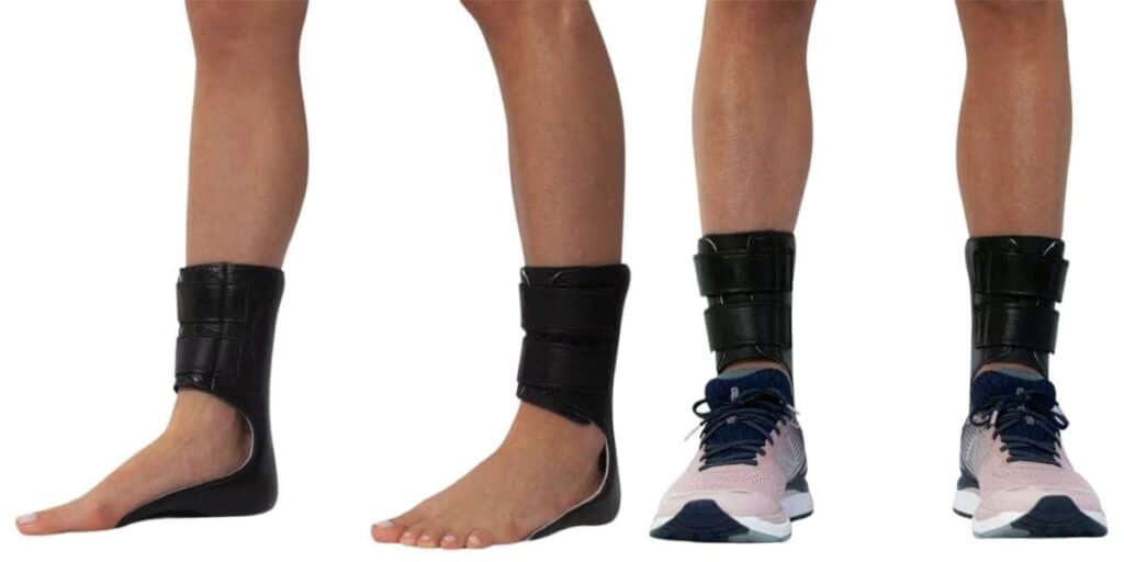
Rarely will I consider the use of ankle-foot orthoses, as I find young adults and teens do not tolerate them well. I primarily use them in nonoperative cases, mainly for either severe deformity or athletes. Again, proper posting of the orthotic portion and cushioning of the medial malleolus and navicular regions will make for a more comfortable device, based on my observations.
When Pediatric Flatfoot Requires Surgery
My number 1 go-to procedure in pediatric flatfoot correction is an Evans calcaneal osteotomy (Figure 2). I find the procedure to be exceptionally strong in the amount of correction it offers; it can be performed at a young age, as there is no growth plate in the region, and it does not increase forefoot varus during correction. Although subtalar implants are gaining popularity and are relatively easy to perform, I have found them difficult for patients to tolerate. In my observation, the varus correction of the hindfoot often increases forefoot deformity. With an Evans procedure, one can correct the heel valgus, along with the tall navicular unroofing and lateral deviation at the midtarsal joint.
Finally, by adding stretch to the peroneus longus tendon with an Evans procedure, there is a plantarflexion of the first metatarsal. All of these deformities are critical to correct in flexible flatfoot, which is why I find the Evans procedure to be the single most powerful option for deformity correction.
I treat the forefoot in two different ways. In very hypermobile deformities, I will look for a naviculocuneiform sag or a hypermobile first ray. If there is a naviculocuneiform sag, I will fuse that joint. If there is a very hypermobile first ray, I will perform a first metatarsocuneiform fusion. However, in most cases, I find the Evans procedure will correct some of the laxity as the peroneus longus is put on stretch, and that correction of the forefoot varus is often possible with a Cotton cuneiform osteotomy and plantarflexion of the first metatarsal and ray. It is important to consider the options prior to surgery, and if there is doubt, in my experience, fusion is often a better option.
I have started to shy away from standard gastrocnemius or Achilles lengthening. If the foot can be brought close to neutral during examination, I will avoid lengthening, as it weakens the muscle/tendon in young patients and may prevent them from returning to active, competitive sports. I will warn the patient and their parents that there may be a need for a lengthening in the future if pain or deformity persists; however, with proper preoperative planning, I have found it rarely necessary to do a second surgery. If a lengthening is necessary, I will lengthen the medial gastrocnemius only and see how much motion I get. This is often enough and leaving the lateral muscle-tendon junction intact keeps some additional strength for the young patient.
Removal of the accessory navicular and imbrication of the posterior tibial tendon are also considerations in pediatric patients. I find this will strengthen the pull on the medial column and will improve the function of the foot. If there is a tear in the spring ligament, this can be a serious and challenging problem to address. There are a few options, such as an internal brace or fusion of the talonavicular joint, that can work well, but my personal option is to perform a repair of the spring ligament directly with anchors into the navicular and imbrication of the spring ligament with an Artelon graft (Artelon). The graft is fairly strong, in my observation, and can be incorporated into the distal posterior tibial tendon to enhance the region’s strength. I have also found that the Evans calcaneal osteotomy can reduce strain on the talonavicular joint and should be considered essential in cases of spring ligament tears.
In Conclusion
With proper planning and a detailed surgical repair of pediatric flexible flatfoot, patients can achieve a wonderful and long-lasting result. It is essential to plan ahead for all three planes of deformity and to consider which surgical procedures will help correct both the primary and secondary deformities. Finally, postoperative gait training and foot and ankle strengthening will help the patient return to full activity and potentially even high-level competitive sports after surgery.
Dr. Baravarian is an Assistant Clinical Professor at the UCLA School of Medicine.