Locations
Dr. Baravarian discusses tarsal tunnel syndrome's causes, symptoms, and treatment options
What's tarsal tunnel syndrome?
The tarsal tunnel is located inside the ankle. Running next to the ankle bones, this narrow tunnel serves as a pathway for many of the foot and ankle’s tendons, nerves, and blood vessels. It protects many vital structures, such as the posterior tibial nerve, and maintains foot strength, flexibility, and health.
Tarsal tunnel syndrome (TTS) is a disorder caused by trauma to the tibial nerve (or its branches). Similar to carpal tunnel syndrome of the wrist, it is usually due to compression or entrapment of the nerve as it passes through the tarsal tunnel.
What causes tarsal tunnel syndrome?
Tarsal tunnel syndrome is a form of neuropathy (nerve damage) that results from the squeezing of the tibial nerve. Common causes of tarsal tunnel syndrome include other conditions of the feet and ankles, such as:
- Flat feet can stretch the tibial nerve.
- Bony growths in the tarsal tunnel.
- Varicose veins surrounding the tibial nerve can cause nerve compression.
- Inflammation from rheumatoid arthritis.
- Lesions and masses such as ganglion cysts or tumors near the tibial nerve.
- Inflammation and swelling from an ankle sprain or fracture.
- Diabetes makes the nerves more vulnerable to compression.
What are the symptoms of tarsal tunnel syndrome?
Many patients with TTS experience a tingling or burning sensation along the tarsal tunnel. Discomfort can run from the inside of the ankle down to the arch of the foot. While pain is often limited to the ankle and the bottom of the foot, other individuals can experience heel pain and pain in their toes. The severity and sensation of foot pain associated with TTS can range from numbness to burning, tingling, or shooting pain akin to an electrical shock.
It is crucial to seek attention from healthcare providers at the onset of symptoms to diagnose or rule out tarsal tunnel syndrome. When caught and treated early, Patients have many conservative treatment options. However, if the condition is left to progress untreated, it can result in permanent nerve damage.
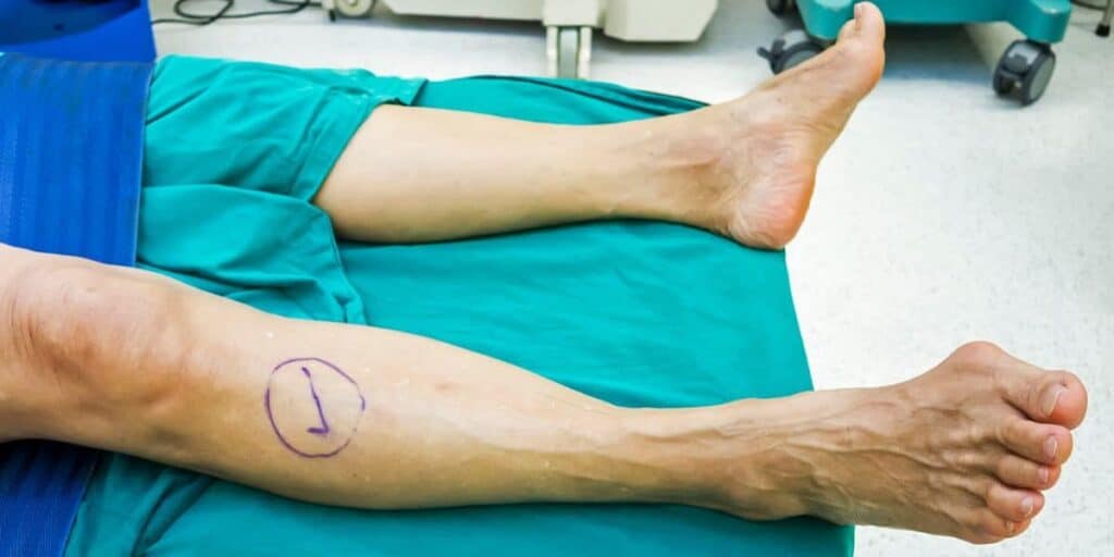
How does Dr. Baravarian diagnose tarsal tunnel syndrome?
Dr. Baravarian has access to nerve conduction studies, ultrasound, X-rays, and MRIs to diagnose the extent of your nerve compression.
A nerve conduction study is typically used in cases of neuralgia to ensure that a peripheral nerve, such as those in the tarsal tunnel, is functioning correctly. Mild electrical stimulation is provided to the nerve during the procedure, known as electromyography (EMG). Electrodes, placed at varying points along the nerve, assess nerve function by recording the speed and strength of the current as well as the resulting muscle movement.
The test is different than imaging tests like an X-ray or MRI, which show if any of the soft tissue, bones, or ligaments are impinging on the space occupying the peripheral nerves.
In some cases, electrical stimulation may not be needed. Dr. Baravarian may use Tinel’s test, in which he manually taps on the tarsal tunnel. If this action causes pain or tingling, it is a Tinel sign that TTS is likely present. Similarly, dorsiflexion eversion (pushing the ball of the foot toward the shin), which can produce symptoms, may also indicate TTS.
Dr. Bob occasionally sees patients who have been previously misdiagnosed with plantar fasciitis or bone spurs. He has decades of combined experience and access to the latest tarsal tunnel syndrome testing equipment for the most accurate diagnosis.
What are conservative treatments for tarsal tunnel syndrome?
If compression is minimal, nonsurgical treatment can be attempted, such as:
- Physical therapy
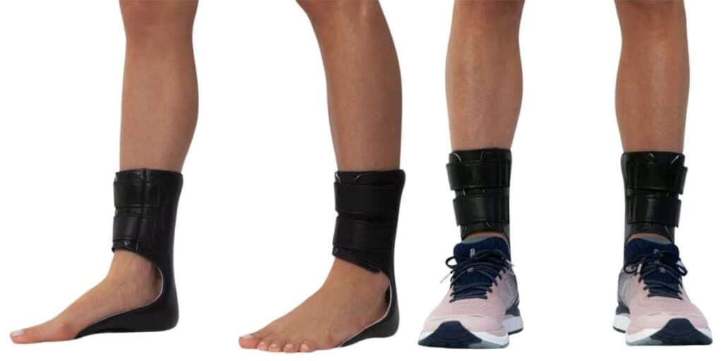
- Custom orthotics, orthopedic braces, and splints
- Rest
- Casting and immobilization
- Nonsteroidal anti-inflammatory medications (NSAIDs)
- Steroid injections (corticosteroid injections)
If these less-invasive methods fail to resolve the compression, Dr. Baravarian may need to consider surgical intervention.
Tarsal tunnel syndrome surgery
A tarsal tunnel syndrome surgical procedure should only be considered if all non-operative treatment options have been unsuccessful at relieving pain and providing for decompression of the nerve.
To decompress the nerve in the tarsal tunnel, Dr. Baravarian makes an incision behind the ankle bone and toward the bottom of the foot. The posterior tibial nerve is separated from the artery and vein and then follows into the tunnel, often to its narrowest point, where it meets the abductor hallucis muscle toward the bottom of your foot. Here, it splits into two primary branches: the medial plantar and the lateral plantar nerve. If either is affected, it could lead to heel or toe pain, respectively. The nerves are then released from impingement.
Dr. Bob will also look for cysts and other nerve blockage problems that may need correction. If there is scarring within the nerve or branches, the outer layer of nerve wrapping is opened, and scar tissue is removed.
Dr. Baravarian utilizes the state-of-the-art Medtronic intraoperative nerve-testing machine during tarsal tunnel release. Testing ensures that compression issues are identified and remedied. This intra-operative testing is only available in select locations in the United States, including Dr. Baravarian’s locations.
What to expect after tarsal tunnel syndrome surgery
During ankle surgery, a bulky dressing is applied to the foot to keep the leg from moving and minimize swelling during weight-bearing. The dressing typically remains on for about a week. Most people use crutches for the first three weeks and have to limit physical activity to allow the foot to heal.
Sutures are left in place for three weeks until the skin has regained 90% of its original strength. You’ll want the nerves to glide post-operatively, so it is critical during the second and third weeks following the surgery that the posterior tibial nerve and its branches can move freely in the tunnels. To facilitate this, an air cast is used instead of a rigid cast, as it allows for some range of motion and movement of the toes, so the nerves do not adhere to the surrounding tissue.
Why Dr. Baravarian is the best choice for foot and ankle care
Accurate diagnosis of tarsal tunnel syndrome is crucial to stopping further damage. With decades of experience armed with state-of-the-art testing techniques, Dr. Bob can ensure a pinpoint diagnosis.
Dr. Baravarianis an internationally recognized foot and ankle specialist. He uses the latest technologies and treatment options available in a comfortable and family-friendly environment. New patients or individuals concerned about foot or ankle symptoms in the greater Los Angeles area are encouraged to call or schedule a consultation; please call (855) 557-5400 or make an appointment now.
Dr. Bob Baravarian is conveniently located in Los Angeles, near Cedars-Sinai Medical Center, providing expert foot and ankle care for patients throughout Southern California.
Tarsal tunnel syndrome FAQs
How long does tarsal tunnel syndrome last?

Tarsal tunnel syndrome cannot go away on its own. Symptoms can be managed with therapy and medication. Curing tarsal tunnel syndrome involves treating the underlying condition and decompressing the nerve.
Can you have carpal tunnel and tarsal tunnel at the same time?
Carpal tunnel syndrome and tarsal tunnel syndrome are two distinct conditions, with the former affecting the wrist and hand, and the latter involving the ankle and foot. However, research has shown that individuals prone to a narrow carpal tunnel may be more likely to have a narrow tarsal tunnel, increasing the likelihood of developing both conditions, potentially simultaneously.
What can be mistaken for tarsal tunnel syndrome?
Tarsal tunnel syndrome symptoms can sometimes be misdiagnosed, instead being attributed to plantar fasciitis, Achilles tendonitis, or a heel spur (calcaneal spur).
Sources
Tarsal tunnel syndrome
https://doi.org/10.1016/j.foot.2015.08.008Tarsal Tunnel Syndrome: A Review of the Literature
https://doi.org/10.1177/107110079902000312 MRImaging of Entrapment Neuropathies of the Lower Extremity
https://doi.org/10.1148/rg.304095188