The talus bone is commonly referred to as your ankle bone, sitting at the base of your tibia (shin bone) and fibula (calf bone). It helps support the weight of your leg during activities like walking and allows your foot a range of motion for both upward and downward movement.
However, unlike most bones, no muscles attach to the talus; its position relies on the surrounding bones. The talar dome consists of three parts: the talus head, body, and neck. The top of the talar dome, known as the talus body, is entirely covered by cartilage, giving it a rounded dome shape.
Any part of the talar dome can be damaged along with other ankle injuries, and Dr. Bob will often examine each area to diagnose your ankle pain. One such injury is chondral damage, sometimes referred to as a talar dome lesion or osteochondral defect (OCD).
It occurs when damage happens to the articular cartilage atop the talus dome and the subchondral bone below.
What causes a talar dome lesion?
Osteochondral lesions of the talus (OLTs) most commonly occur due to a traumatic injury, such as an ankle sprain, where the foot and ankle joint are twisted or compressed. In some instances, the bones in the ankle joint collide, damaging the surface of the overlying cartilage and resulting in an osteochondral fracture.
If cartilage heals improperly, blood flow to the underlying bone may be interrupted, leading to osteochondritis dissecans (OCD), a condition where bone and cartilage can break off and float in the ankle. This can lead to chronic pain and ankle weakness.
However, other causes of osteochondral lesions of the talar dome include:
- Avascular necrosis (poor blood supply to the bone results in bone death)
- Repetitive motion and stress on the joint
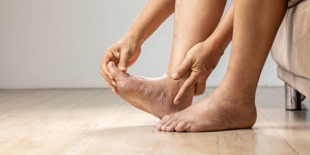
What are the symptoms of talar dome lesions?
In most cases, symptoms of a talar dome injury will appear after the symptoms of the initial ankle injury have eased. The symptoms will vary depending on the location of the injury (medial, posterior, or anterior side of the dome).
Patients with talar dome lesions may experience:
- Pain that resolves after the initial trauma but then returns
- Pain that worsens with activity
- Swelling, ankle instability, and locking of the ankle joint
- The feeling that the ankle joint clicks or catches
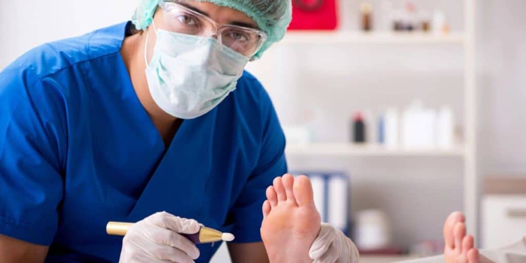
Diagnosing an osteochondral lesion of the talus
Dr. Bob Baravarian will start by asking about your medical history, including any recent ankle fractures or sprains. He will also conduct a physical exam, examining and moving your ankle to assess your range of motion and identify the injured area.
Dr. Bob’s clinics also feature X-ray, CT scan, and MRI machines. X-rays enable him to examine the bones of the ankle joint, while MRIs and CT scans reveal the soft tissue, including ligaments, tendons, and cartilage.
Many times, talar dome lesions can be identified visually.
What are the treatment options for talar dome lesions?
Dr. Bob is among the world’s leading foot and ankle surgeons. He is renowned for treating large osteochondral lesions of the talus through cartilage removal or transplant.
However, when possible, Dr. Bob will try to use a non-surgical approach first.
Non-surgical treatments for talar dome lesions
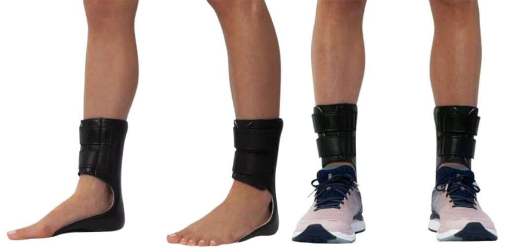
For mild cases of talar dome lesions, where there are no floating pieces of bone or cartilage, Dr. Bob may recommend the following:
- Immobilization of the ankle joint with a non-weight-bearing boot or cast.
- Physical therapy aims to restore the range of motion after the lesion has healed.
- Nonsteroidal anti-inflammatory drugs (NSAIDs) to help with inflammation and pain.
- Amniotic stem cell and platelet-rich plasma injections can be used to help repair damaged cartilage by providing cells and signals that enable the body to heal the cartilage on its own.
- Shockwave or Softwave therapy can be used to aid in the healing of damaged cartilage by signaling the body to repair the affected tissue.
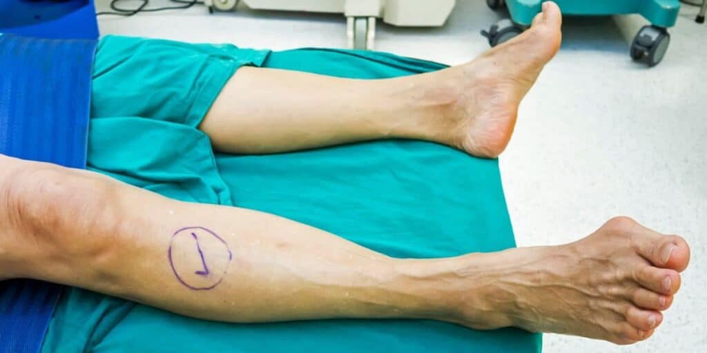
Surgical talar dome lesion treatment
Talar dome lesions are classified into stages using the Berndt and Harty classification system, which ranks injuries from stage I to stage IV. Treatment options, depending on the extent of cartilage damage, may include arthroscopic removal of the damaged cartilage and drilling of the affected area.
After arthroscopic debridement, in which Dr. Bob cleans the joint by removing debris and relieving any impingement, Dr. Bob will perform arthroscopic bone marrow stimulation. During this minimally invasive surgical technique, he causes microfractures in the anterolateral and mid-lateral areas of the talus. These openings allow blood and bone marrow to enter the damaged tissues, promoting the development of new cartilage (fibrocartilage).
However, if the lesion is large or affects the deep bone beneath the cartilage-damaged area, a different type of surgical treatment may be necessary. Dr. Bob might perform a transplant of fresh bone and cartilage. The bone can be harvested from the patient’s own body, typically in the form of a tibial autograft, or it may come from a lab, known as an allograft.
Autologous grafts using tissue from your own body typically have a lower risk of complications, as there is almost no chance of your body rejecting its own tissue.
Dr. Bob implants the bone graft into the injured area, addressing the problematic regions and promoting healing. The frequency of bone grafting is significantly lower than that of arthroscopy clean-out and bone marrow stimulation, as it is reserved for only the most severe cases.
BioCartilage transplant for osteochondral lesion treatment
BioCartilage is a groundbreaking product that leads to a stronger ankle joint, quicker recovery, and a lower risk of re-injury compared to traditional methods. Read more about BioCartilage.
What to expect after talar dome osteochondral lesion surgery
Because the talus bone has a limited blood supply, healing a broken talus can take longer than healing most bones, with recovery from talar dome lesion surgery often requiring months.
Patients recovering from a talar dome osteochondral lesion treatment may not be able to walk without crutches for several weeks, after which they will likely need to wear a walking cast or ankle brace for several additional months.
During gradual rehabilitation, we’ll recommend pain management medications and gentle exercises, usually through physical therapy, to help you regain strength, stability, and range of motion in your ankle. Following Dr. Bob’s advice throughout the entire healing process is crucial, as doing too much too soon can risk reinjury or delay recovery.
Dr. Bob has expertise in talar dome lesions.
Dr. Bob has decades of experience treating all forms of ankle injuries and concerns. He utilizes the latest technologies to successfully diagnose and treat talar dome lesions in the most minimally invasive manner possible. New patients or those worried about foot or toe pain in the greater Los Angeles area are encouraged to call or schedule a consultation.
Dr. Bob Baravarian is conveniently located throughout Southern California and the Los Angeles area, including regions in or near Santa Monica, Sherman Oaks, Beverly Hills, West Los Angeles, the San Fernando Valley, El Segundo, the South Bay, LAX, Calabasas, Agoura Hills, and Downtown Los Angeles.
Osteochondral Lesion FAQs
What is a talar dome fracture?
A talar dome fracture is sometimes used interchangeably with the term talar dome lesion. However, a lesion refers to the tearing or fracturing of the talar dome. In contrast, a talar dome fracture generally describes the condition when a piece of cartilage breaks off from the talar dome.
Does a talar dome lesion need surgery?
If a talar dome injury is detected early enough, non-surgical treatment may alleviate symptoms and facilitate healing in the area. However, surgery for talar dome lesions is often necessary if cartilage pieces have broken off and migrated into the joint, or if conservative treatment has failed to manage symptoms.
Will a talar dome lesion go away on its own?
Typically, talar dome injuries will not resolve on their own without intervention. Immobilization and rest may allow minor cases to heal. Meanwhile, other talar dome lesions may require surgical cleaning, micro-fracturing to promote healing, or bone grafting.