Locations
Diego came to UFAI after falling twenty feet from scaffolding. Here's his story.
What's a calcaneus (heel bone) fracture?
A calcaneus fracture is a break in the large heel bone of your foot.
Calcaneal fractures are serious injuries typically seen in patients who have experienced a high-energy event causing axial loading of the bone, where stress is applied along the bone’s axis. Common mechanisms of injury include landing on your feet after a long fall or being in the front seat during a car accident.
What are the types of calcaneal fractures?
Traumas that cause a calcaneus fracture can also harm other tissues. Some of these injuries are more severe or harder to treat than others.
The main types of calcaneal fracture include:
Intra-articular fractures
These involve damage to the cartilage between the joints and are considered the most serious type of heel fracture.
Avulsion fractures
This foot fracture includes a sliver of bone splitting off from the calcaneus due to pulling from the Achilles tendon or another ligament.
Multiple fracture fragments
This type of fracture is also known as a crushed heel injury.
Stress fractures
While most calcaneal fractures are caused by trauma, they can also occur from overuse.
Open fractures
Open fractures involve a break in the bone that is exposed through a wound in the skin. They are also known as compound fractures.
What are the symptoms of a calcaneal fracture?
The calcaneus (heel bone) and the talus (the lowest bone of the ankle) form the subtalar joint—an essential part of biomechanics involved in flexing, standing, and walking. The calcaneus also creates a joint with the cuboid bones. Five ligaments connect the calcaneocuboid joint, making it another important joint for movement.
Because the calcaneus is part of these important joints, an injury to the bone can cause many problematic symptoms.
- Sharp, severe heel pain
- Swelling and bruising in the heel
- Inability to bear weight on the affected foot
- General pain in the heel that gradually worsens
- Blisters may develop around the heel if the fracture causes swelling
How is a calcaneal fracture diagnosed?
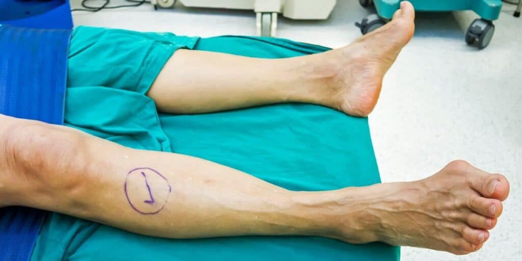
The first step in diagnosing a calcaneal fracture is a physical exam by Dr. Bob Baravarian, a foot and ankle specialist. Dr. Bob will assess your foot for swelling, range of motion, and other signs of joint damage.
X-ray imaging can assist in evaluating the injury by visualizing the bone and calculating Bohler’s angle. Once Dr. Bob has determined the extent of the injury, he can choose the best treatment option. For improved visualization of the joint, Dr. Bob has access to CT scans to view the fracture pattern and assess whether surgery is necessary.
Depending on the cause of your injury, Dr. Bob may also examine you for an ankle or mid-foot injury or refer you to another specialist to assess injuries beyond the foot. About 10% of patients with calcaneal fractures also experience a back injury, which occurs when a vertebra in the lower-mid back is crushed.
What are the calcaneal fracture treatment options?
Heel bone fractures are notoriously hard to treat and usually take a long time to heal. Fixing a calcaneal fracture is challenging because the break is rarely a clean break like you might see in a broken shin or arm.
Think of the calcaneus like a hard-boiled egg: its outer layer is tough and brittle, while the inner tissue is soft and spongy. When you crack a hard-boiled egg, the shell breaks apart. Similarly, when the calcaneus fractures, its hard outer layer can break into irregular fragments.
Non-surgical treatment
A conservative, non-invasive treatment plan may be suggested for patients who are not suitable candidates for surgery. Smokers, older adults, and patients with diabetes or vascular disease might face a higher risk of surgical complications, such as infection or blood loss.
Although calcaneal fractures can be quite severe, studies have indicated that nonoperative treatment can be nearly as effective as surgery. Usually, non-surgical approaches include:
- About 10-12 weeks of non-weight bearing.
- Immobilization in a cast or splint for one to two weeks.
- Lifting the foot to or above heart level.
- Apply ice to the heel for 10-15 minutes, three times a day, to reduce swelling and pain.
- A compression sock or bandage to hold the joint and reduce swelling.
- Depending on the pain level, over-the-counter NSAIDs like ibuprofen or prescription pain medication can help reduce pain as you heal.
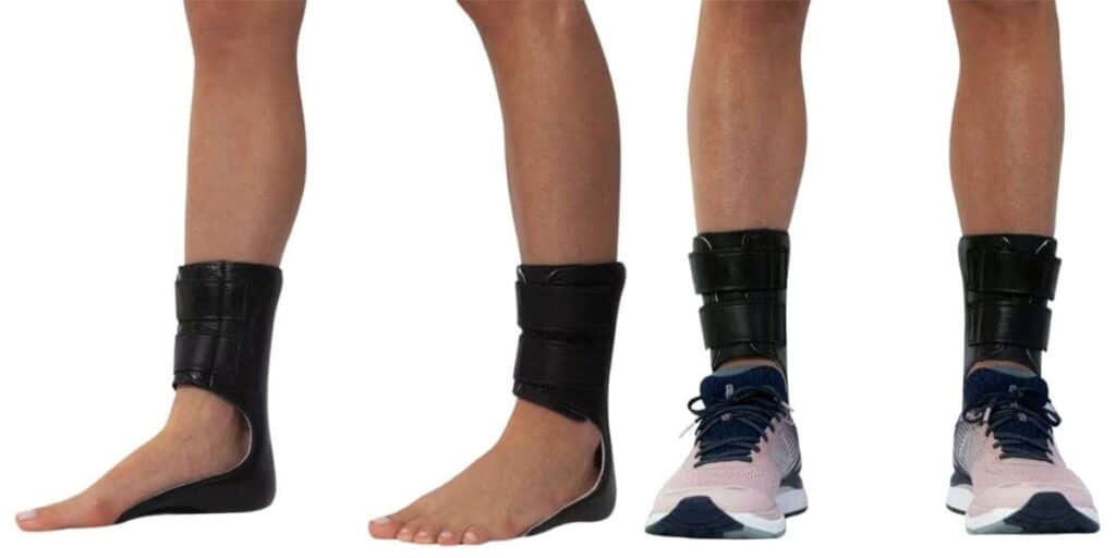
- After two weeks of healing, you can start physical therapy exercises to regain your range of motion.
Surgical treatment
Surgical correction of a calcaneal fracture requires an extremely skilled surgeon and carries inherent risks. Dr. Bob will reconstruct the heel bone to closely resemble its original shape. Since each fracture is different, every surgical procedure is highly tailored.
Surgery usually cannot start until the swelling decreases, which typically takes about 10-14 days after the injury. Operating on a foot that is too swollen can cause healing issues and raise the risk of infection.
The surgery is usually done through an open cut on the outer side of the heel. Dr. Bob then carefully realigns the fragments and secures them in place with screws and metal plates.
Percutaneous treatment
This minimally invasive surgical method can be used on fewer than 10% of fractures. The surgeon makes a small incision and threads a surgical wire through the broken bone pieces. The surgeon can then realign the bone fragments into the proper position.
What are the possible complications of calcaneal fracture surgery?
Surgical treatment of a fractured heel bone carries risks. Common complications include:
- Infection. Osteomyelitis is a serious deep wound infection that affects the bone, and the calcaneus is especially vulnerable. In many cases, a bone infection requires treatment with amputation.
- Wound complications: Since circulation to the soft tissues of the heel is relatively poor, the surgical site might not heal properly.
- Subtalar arthritis is a long-term pain condition that often affects people with healed calcaneal fractures.
- Nerve damage
- The inability of the bone to mend
- Compartment syndrome
What is the recovery from calcaneal fracture surgery?
After surgery, Dr. Bob will probably immobilize your foot in a cast to help the bone and joints heal. Follow the at-home treatments advised for non-surgical patients, including rest, ice, elevation, and compression.
You should avoid putting weight on your foot for 10-12 weeks to allow the heel to heal properly. At two weeks, your specialist may remove the cast and recommend physical therapy exercises.
Dr. Bob is a trained podiatric trauma specialist with decades of experience treating foot and ankle fractures. They provide advanced care in a compassionate, relaxed environment with some of the highest success rates in the country.
Why Dr. Bob Baravarian is your best choice for foot and ankle care in Los Angeles
Using the most advanced techniques, some developed with Dr. Bob’s help, has enabled him to achieve some of the highest success rates in the country for ankle injuries. His goal is to get you back on your feet quickly, using the least invasive treatments possible.
Patients are his top priority. From the simplicity of scheduling your appointment, Dr. Bob’s family-friendly office staff supports you every step.
Podiatric foot and ankle surgeons focus exclusively on the foot and ankle from the start of medical school. After completing their medical training, they undertake a rigorous three-year surgical residency. What sets podiatric surgical residents apart from general orthopaedic surgeons is that they specialize in the foot and ankle, whereas most (though not all) orthopaedic residents do not.
Years of training, decades of experience, and research are why Dr. Bob maintains among the highest success rates in the United States, genuinely helping thousands get back on their feet and return to their lives.
Dr. Bob Baravarian is conveniently located in Los Angeles, near Cedars-Sinai Medical Center, providing expert foot and ankle care for patients throughout Southern California.
Calcaneal fracture FAQs
What are extra-articular fractures?

Extra-articular fractures occur outside the joint space, meaning they do not directly involve the joint surfaces. This contrasts with intra-articular fracture lines, which happen within the joint space and can disrupt the joint surface.
How common are calcaneal fractures?
Seven bones — called tarsals — form the hindfoot and midfoot. The calcaneus is the largest of these tarsal bones. Fractures of the tarsal bones make up about 2% of all adult fractures, and only half of tarsal fractures involve the calcaneus.
Sources
Adams MR, Koury KL, Mistry JB, Braaksma W, Hwang JS, Firoozabadi R. Plantar Medial Avulsion Fragment Associated With Tongue-Type Calcaneus Fractures. Foot Ankle Int.
https://pubmed.ncbi.nlm.nih.gov/30841752/Renfrew DL, el-Khoury GY. Anterior process fractures of the calcaneus. Skeletal Radiol
https://pubmed.ncbi.nlm.nih.gov/4023740/Shih JT, Kuo CL, Yeh TT, Shen HC, Pan RY, Wu CC. Modified Essex-Lopresti procedure with percutaneous calcaneoplasty for comminuted intra-articular calcaneal fractures: a retrospective case analysis. BMC Musculoskelet Disord.
https://pubmed.ncbi.nlm.nih.gov/29523122/Siebert CH, Hansen M, Wolter D. Follow-up evaluation of open intra-articular fractures of the calcaneus. Arch Orthop Trauma Surg. 1998;117(8):442-7.
https://pubmed.ncbi.nlm.nih.gov/9801778/Seat A, Seat C. Lateral Extensile Approach Versus Minimal Incision Approach for Open Reduction and Internal Fixation of Displaced Intra-articular Calcaneal Fractures: A Meta-analysis. J Foot Ankle Surg. 2020 Mar-Apr;59(2):356-366.
https://pubmed.ncbi.nlm.nih.gov/32131003/Jiménez-Almonte JH, King JD, Luo TD, Aneja A, Moghadamian E. Classifications in Brief: Sanders Classification of Intraarticular Fractures of the Calcaneus. Clin Orthop Relat Res.
https://pubmed.ncbi.nlm.nih.gov/30664605/